
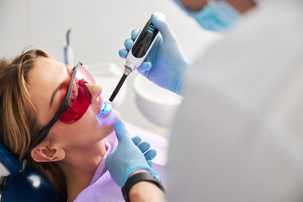
Dental fillings serve as more than simple cavity repairs. These restorations establish antimicrobial barriers that halt bacterial progression into vulnerable dentin and pulp tissues. Modern adhesive materials create hermetic seals that prevent microleakage while stabilizing compromised tooth structure. The biomechanical reinforcement provided by contemporary filling materials addresses multiple failure mechanisms simultaneously. Understanding these protective mechanisms reveals why prompt restoration remains fundamental to preserving natural dentition and avoiding complex endodontic interventions.
The Barrier Effect: How Fillings Block Bacterial Invasion
A dental filling creates a physical seal that prevents oral bacteria from penetrating compromised tooth structure. This barrier eliminates the protected environment where pathogenic microorganisms proliferate within cavitated lesions. By occluding access points, the restoration disrupts established bacterial colonies and prevents new colonization of dentinal tubules.
The filling material achieves biofilm disruption by removing the retentive surfaces that harbor cariogenic bacteria. Modern restorative materials bond intimately to tooth structure, creating a hermetic seal at the tooth-restoration interface. This prevents microleakage and subsequent bacterial infiltration that would otherwise compromise pulpal health.
Additionally, certain filling materials facilitate remineralization enhancement through fluoride release and ion exchange mechanisms. Glass ionomer and resin-modified materials actively contribute to the repair of demineralized tooth structure adjacent to the restoration, strengthening the remaining tooth and providing ongoing protection against secondary caries development.
Stopping Decay in Its Tracks: The Protective Seal of Modern Fillings
The protective seal established by contemporary dental restorations halts caries progression through mechanical isolation of affected dentin and enamel from the oral environment. Modern composite resins and glass ionomers create hermetic barriers that prevent substrate demineralization while eliminating bacterial nutrient access. This occlusive interface disrupts acidogenic biofilm metabolism, effectively arresting lesion advancement.
Clinical studies demonstrate 95% caries arrest rates when restorations maintain marginal integrity. The seal’s efficacy depends on proper cavity preparation, moisture control during placement, and material polymerization protocols. Patients must implement improved oral hygiene techniques post-restoration to prevent secondary caries formation at restoration margins. Timely dental checkups enable early detection of microleakage or material degradation. Radiographic assessment during routine examinations identifies recurrent decay beneath existing restorations, facilitating prompt intervention before pulpal involvement occurs.
Structural Reinforcement: Restoring Your Tooth’s Natural Strength
When dental caries compromises tooth structure, modern restorative materials provide biomechanical reinforcement that approximates original dentinal properties and enamel characteristics. Contemporary filling materials restore biomechanical stability through adhesive bonding mechanisms that integrate seamlessly with remaining healthy tissue. This integration distributes masticatory forces uniformly across the restored surface, preventing stress concentration points that typically lead to fracture propagation.
Advanced composite resins and amalgam formulations achieve tooth reinforcement by replacing lost dentin and enamel with materials exhibiting comparable elastic moduli. The restorative process eliminates unsupported cusps and undermined areas susceptible to breakage during normal function. Properly contoured fillings reestablish ideal occlusal relationships, ensuring balanced force distribution during mastication. This structural rehabilitation prevents progressive weakening that occurs when cavitated teeth remain untreated, ultimately preserving long-term dental integrity and functionality.
Preventing Root Canal Infections Through Early Intervention
Most untreated dental caries progress through dentin toward pulpal tissue, establishing pathways for bacterial invasion that necessitate endodontic therapy. Early diagnosis identifies cavities before bacterial penetration reaches the pulp chamber, where infection triggers irreversible pulpitis. Radiographic examination and clinical assessment detect enamel breaches and dentinal involvement, enabling prompt intervention.
Proactive treatment through conservative restorations eliminates cariogenic bacteria and seals dentinal tubules against microbial colonization. Placement of appropriate filling materials creates antimicrobial barriers while preserving essential pulp tissue. Without intervention, bacterial toxins compromise pulpal vasculature, causing necrosis and periapical pathology.
Studies demonstrate that teeth receiving timely restorations exhibit markedly lower endodontic treatment rates compared to those with delayed intervention. Prompt filling placement maintains pulpal health, preventing complications requiring root canal therapy, apicoectomy, or extraction.
Maintaining Proper Bite Alignment and Jaw Function
Although occlusal surfaces deteriorate gradually from carious lesions, untreated cavities create irregular tooth morphology that disrupts masticatory biomechanics and temporomandibular joint function. Dental caries progressively destroy enamel and dentin, forming concavities that alter occlusal plane relationships. These structural deficiencies redistribute occlusal forces unevenly across remaining tooth structure, potentially causing premature wear of opposing dentition.
Restorative procedures restore anatomical contours through proper occlusal adjustments, reestablishing harmonious intercuspation patterns. Clinicians meticulously shape filling materials to replicate natural cusp heights and fossae depths, ensuring optimized masticatory function. Precise contouring prevents hyperocclusion or infraocclusion, maintaining physiological vertical dimension. Contemporary composite materials enable accurate reproduction of occlusal anatomy, preserving proprioceptive feedback mechanisms essential for neuromuscular coordination. Without timely intervention, malocclusion secondary to tooth structure loss precipitates muscular dysfunction, articular disc displacement, and degenerative joint changes.
Different Filling Materials and Their Protective Properties
The selection of restorative filling materials directly influences the tooth’s long-term structural integrity and resistance to secondary decay. Composite resin fillings provide superior adhesive bonding to tooth structure, effectively sealing margins against bacterial infiltration while preserving maximum healthy tooth tissue through conservative preparation techniques. Amalgam restorations demonstrate exceptional compressive strength and wear resistance in posterior teeth, maintaining occlusal stability for decades when properly placed and contoured.
Composite Resin Benefits
When selecting restorative materials for cavity treatment, composite resin offers distinct protective advantages that extend beyond basic structural repair. The material’s chemical bonding capability creates a microscopic seal with tooth structure, effectively preventing bacterial infiltration and secondary caries formation. This adhesive property eliminates microleakage pathways that commonly compromise restoration longevity.
Beyond functional protection, composite resin’s aesthetic appeal preserves natural tooth appearance through customizable shade matching, maintaining patient confidence while ensuring ideal oral health. The material’s durability attributes include resistance to moderate occlusal forces and thermal expansion coefficients similar to natural dentition, minimizing stress-induced fractures. Composite resin polymerization creates a hardened barrier that withstands acidic environments, providing long-term defense against demineralization. These combined properties establish composite resin as a comprehensive preventive solution for arresting progressive tooth destruction.
Amalgam Durability Features
Material selection for cavity restoration requires evaluation of multiple protective factors, with amalgam presenting distinct durability characteristics that differ fundamentally from composite systems. The metal composition of dental amalgam—primarily silver, tin, copper, and mercury—creates an alloy with exceptional mechanical strength and longevity. This formulation demonstrates superior corrosion resistance in the oral environment, maintaining structural integrity despite constant exposure to saliva, temperature fluctuations, and pH variations.
Amalgam’s protective capabilities extend beyond initial placement. The material exhibits self-sealing properties through gradual corrosion product formation at tooth-restoration interfaces, reducing microleakage over time. Clinical studies document average functional lifespans exceeding fifteen years, with minimal degradation under occlusal forces. The material’s compressive strength prevents fracture propagation in posterior teeth, while its thermal expansion coefficient closely matches natural tooth structure, minimizing stress-induced damage.
The Long-Term Cost Benefits of Timely Filling Placement
Early intervention through filling placement demonstrates significant economic advantages by preventing the progression of carious lesions that would otherwise require endodontic treatment, crown restoration, or extraction with subsequent prosthetic replacement. Most dental insurance plans provide higher reimbursement percentages for preventive and basic restorative procedures compared to major restorative work, with typical coverage ranging from 80-100% for fillings versus 50% for crowns or bridges. The financial differential between a single filling procedure and complex treatment modalities can exceed tenfold, making prompt restoration a cost-effective strategy for maintaining dental health.
Avoiding Complex Procedures
Although dental caries may initially present as minor surface lesions requiring simple restorative treatment, untreated cavities progressively destroy tooth structure and necessitate increasingly invasive interventions. Timely placement of amalgam or composite restorations prevents bacterial infiltration into dentin and pulpal tissues, thereby avoiding costly treatments such as endodontic therapy, post-and-core buildups, and full-coverage crowns.
Research demonstrates that teeth receiving early occlusal fillings exhibit 85% lower rates of pulpal involvement compared to untreated carious lesions over five-year periods. This preventive approach effectively stops demineralization processes while minimizing future work requirements. Each millimeter of preserved tooth structure reduces the probability of catastrophic failure requiring extraction and implant placement by approximately 12%. Strategic intervention at the incipient caries stage maintains structural integrity, preserves natural dentition, and eliminates the cascade of complex procedures.
Insurance Coverage Advantages
How greatly do insurance reimbursement patterns favor preventive restorative care over major reconstructive procedures? Most dental plan coverage structures allocate 70-80% reimbursement for basic restorative fillings, while crown placement typically receives 50% coverage with higher deductibles. Insurance policy requirements often mandate pre-authorization for procedures exceeding $1,000, creating administrative delays for complex treatments.
Annual maximum benefits constrain extensive restorative work, yet routine filling placement rarely approaches these limits. Preventive-focused policies recognize amalgam and composite restorations as cost-effective interventions, processing claims within standard review periods. Conversely, root canal therapy, post-core buildups, and full-coverage restorations exhaust benefits rapidly while requiring substantial patient co-payments. This reimbursement disparity incentivizes timely caries management through judicious filling placement, reducing long-term financial burden on both patients and insurance carriers while maintaining ideal dental health outcomes.
Signs You Need a Filling Before Damage Worsens
When tooth decay progresses beyond initial demineralization, specific clinical indicators signal the need for restorative intervention through dental fillings. Persistent tooth sensitivity to temperature variations, particularly when consuming hot or cold substances, indicates compromised enamel integrity requiring immediate evaluation. Visible cavitation, presenting as dark spots or holes on tooth surfaces, represents irreversible structural damage necessitating prompt treatment.
Additional warning signs include localized pain during mastication, food impaction in specific areas, and detectable surface roughness during tongue exploration. Radiographic evidence may reveal interproximal caries invisible to clinical examination. Early detection through regular dental assessments enables conservative restoration approaches, preserving maximum tooth structure. Delaying intervention allows bacterial penetration into deeper dentin layers, potentially compromising pulpal essentiality and requiring more extensive procedures than simple filling placement.
Frequently Asked Questions
How Painful Is Getting a Filling?
Patients typically experience minimal discomfort levels during filling procedures due to local anesthesia administration. Minor sensitivity may occur temporarily. Post procedure care includes avoiding extreme temperatures and maintaining conducive oral hygiene to safeguard successful restoration integration.
How Long Does the Filling Procedure Typically Take?
The filling procedure duration typically ranges from 20-60 minutes, depending on cavity complexity. Tooth preparation requirements include decay removal, cavity shaping, and surface conditioning. Multiple-surface restorations or deeper cavities necessitate extended treatment time for ideal outcomes.
Can I Eat Immediately After Getting a Filling?
Patients should wait until anesthesia subsides before eating. They must avoid hard crunchy foods for 24 hours post-procedure to prevent restoration compromise. Maintain good oral hygiene through gentle brushing to protect the new filling’s integrity.
How Often Do Fillings Need to Be Replaced?
Dental fillings typically require replacement every 5-15 years, depending on material composition and wear patterns. Proper dental hygiene and regular checkups enable early detection of filling deterioration, preventing secondary caries development and structural compromise.
Will My Tooth Be Sensitive After Getting a Filling?
Patients commonly experience increased temperature sensitivity following restoration placement, particularly to cold stimuli. Transient pulpal inflammation and possible gum irritation typically resolve within 2-4 weeks. Persistent symptoms exceeding this timeframe warrant clinical evaluation for potential complications.