
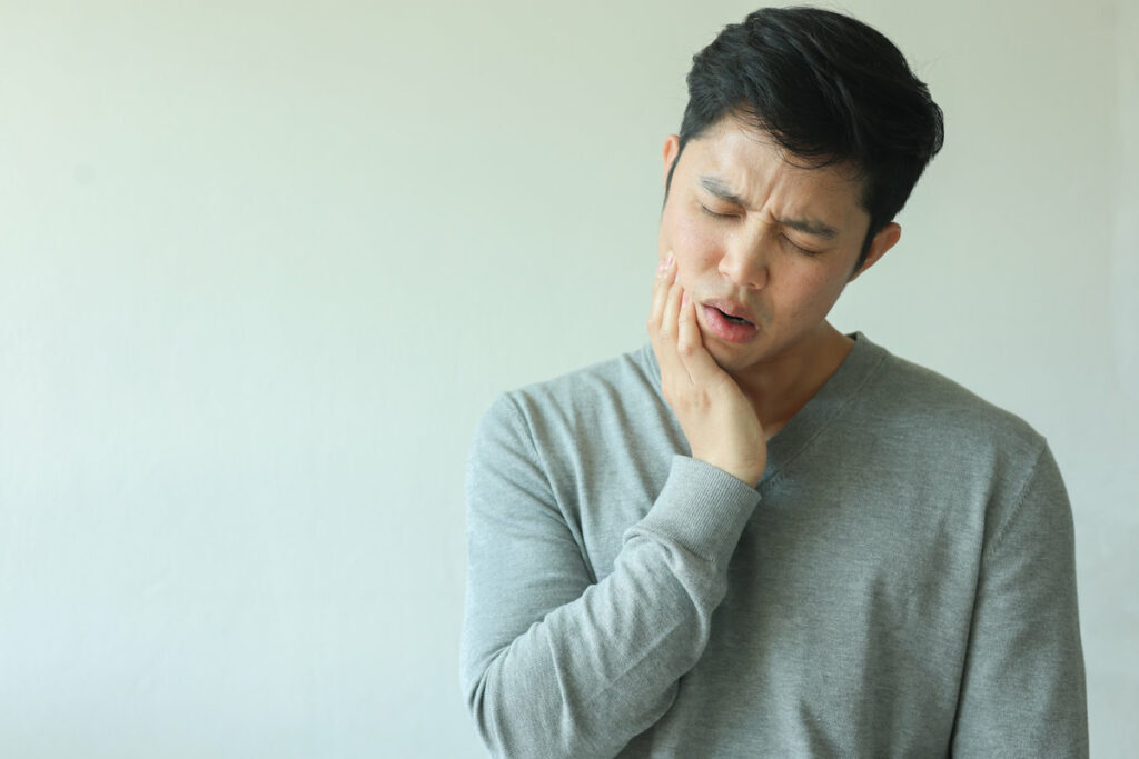
The temporomandibular joint connects the mandible to the temporal bone through a complex articulation involving fibrocartilaginous disc, ligaments, and masticatory muscles. When this intricate system malfunctions, patients experience debilitating orofacial pain, joint sounds, and functional limitations affecting millions worldwide. Current clinical evidence supports multimodal treatment approaches ranging from conservative therapies to surgical interventions. Understanding the pathophysiology and therapeutic options determines successful patient outcomes in managing this prevalent musculoskeletal condition.
Understanding the Temporomandibular Joint and Its Function
The temporomandibular joint (TMJ) represents one of the most complex articulations in the human body, functioning as a bilateral synovial joint that connects the mandibular condyle to the temporal bone’s glenoid fossa. The joint structure comprises an articular disc positioned between these osseous components, dividing the joint space into superior and inferior compartments. This fibrocartilaginous disc enables smooth articulation during mandibular movements.
The joint mechanics involve both rotational and translational movements, allowing for mastication, speech, and facial expression. During mouth opening, the condyle initially rotates within the inferior joint space, followed by anterior translation along the articular eminence. Surrounding ligaments, including the temporomandibular and sphenomandibular ligaments, provide structural stability. The lateral pterygoid muscle directly attaches to the disc-condyle complex, coordinating disc displacement during functional movements.
Common Symptoms of TMJ Disorder
When dysfunction affects this complex articulation system, patients typically present with a constellation of clinical manifestations that vary in severity and presentation. Primary symptoms include myofascial pain affecting the masseter and temporalis muscles, often accompanied by muscle spasms that restrict mandibular range of motion. Articular dysfunction manifests as jaw clicking, crepitus, or reciprocal clicking during condylar translation, indicating disc displacement with or without reduction.
Secondary manifestations encompass otalgia, tinnitus, and cervicogenic headaches resulting from referred pain patterns. Patients frequently report trismus, malocclusion, and deviation of the mandible during opening movements. Chronic cases demonstrate masticatory muscle hypertrophy, dental attrition from parafunctional habits, and temporal summation of nociceptive inputs. Symptom severity correlates with inflammatory mediator levels within the synovial fluid and degree of articular degeneration observed through imaging studies.
Primary Causes and Risk Factors
Temporomandibular joint disorders frequently develop following direct trauma to the mandible or joint capsule, which can result in disc displacement, ligamentous damage, or articular surface disruption. Chronic bruxism and nocturnal teeth clenching generate excessive compressive forces on the TMJ, leading to degenerative changes in the articular cartilage and synovial inflammation. These mechanical stressors account for approximately 70% of TMJ dysfunction cases, with trauma-induced disorders showing higher rates of joint derangement on magnetic resonance imaging.
Jaw Injury Impact
Trauma to the maxillofacial region represents a significant etiological factor in temporomandibular joint dysfunction, with direct impact injuries accounting for 25-50% of TMD cases according to epidemiological studies. Motor vehicle accidents, sports-related trauma, and interpersonal violence commonly precipitate articular disc displacement, capsular ligament disruption, and condylar fractures. Post-traumatic inflammation triggers synovial membrane proliferation and intra-articular adhesion formation, compromising joint biomechanics.
Secondary complications manifest as altered mandibular kinematics, affecting both mastication and articulation. The jaw impact on eating becomes evident through reduced maximum voluntary bite force and asymmetric muscle activation patterns. Similarly, jaw impact on speech presents as impaired consonant production, particularly bilabial and labiodental phonemes. Whiplash-associated disorders induce indirect TMJ trauma through sudden acceleration-deceleration forces, causing retrodiscal tissue compression and anterior disc displacement without reduction.
Teeth Grinding Effects
Beyond traumatic etiology, bruxism emerges as a predominant parafunctional contributor to temporomandibular dysfunction, affecting 8-31% of the general population with higher prevalence in TMD cohorts. Nocturnal teeth grinding patterns generate forces exceeding 250 pounds per square inch, surpassing normal masticatory pressures by 300-600%. These repetitive microtraumas induce articular disc displacement, synovial inflammation, and condylar remodeling.
Excessive teeth clenching during diurnal episodes compounds biomechanical stress through sustained isometric contraction of masticatory muscles, particularly the masseter and temporalis. This chronic hyperactivity triggers myofascial pain syndrome, characterized by trigger points and referred pain patterns. Bruxism-induced enamel attrition alters occlusal vertical dimension, disrupting condyle-fossa relationships and precipitating adaptive changes in joint morphology. Electromyographic studies demonstrate increased muscle activity correlating with TMJ capsular strain and ligamentous laxity.
Diagnosing TMJ Disorder: Tests and Examinations
When patients present with symptoms suggestive of temporomandibular joint dysfunction, clinicians employ a systematic diagnostic approach combining clinical examination with imaging modalities to establish an accurate diagnosis. The clinical evaluation begins with palpation of masticatory muscles, assessment of mandibular range of motion, and auscultation for joint sounds including clicking, popping, or crepitus. Maximum interincisal opening measurements and lateral excursion patterns provide quantitative data regarding joint mobility.
Advanced imaging techniques complement physical findings. Panoramic radiographs reveal gross osseous abnormalities, while magnetic resonance imaging visualizes disc position, morphology, and soft tissue pathology. Computed tomography accurately depicts cortical bone changes, erosions, and osteophyte formation. Cone-beam CT offers three-dimensional joint visualization with reduced radiation exposure. Diagnostic nerve blocks may differentiate arthrogenous from myogenous pain origins, guiding targeted treatment strategies.
Home Remedies and Self-Care Strategies
Conservative management approaches constitute the first-line intervention for temporomandibular joint disorder, with self-directed therapies demonstrating significant efficacy in reducing pain intensity and improving functional outcomes. Heat application to the temporomandibular region for 15-20 minutes promotes vasodilation, reduces muscle tension, and enhances tissue elasticity. Patients should implement dietary modifications, selecting soft-textured foods while avoiding excessive mandibular excursion.
Targeted massage techniques applied to the masseter, temporalis, and lateral pterygoid muscles facilitate myofascial release and improve local circulation. Stress reduction protocols, including diaphragmatic breathing and progressive muscle relaxation, address parafunctional habits contributing to symptom exacerbation. Mandibular rest position maintenance, with teeth slightly separated and tongue positioned against the palate, minimizes joint loading. These evidence-based interventions, when performed consistently, yield measurable improvements in pain scores and mandibular range of motion within 4-6 weeks.
Professional Treatment Options and Medical Interventions
When conservative self-care measures prove insufficient for temporomandibular joint dysfunction, patients may require professional interventions ranging from non-invasive to surgical approaches. Dental splints and occlusal guards function to redistribute masticatory forces and prevent parafunctional habits, while targeted physical therapy protocols address muscular imbalances and restore normal joint biomechanics. Refractory cases may necessitate surgical interventions, including arthrocentesis, arthroscopy, or open joint procedures to correct structural abnormalities of the TMJ complex.
Dental Splints and Guards
Three primary categories of oral appliances serve distinct therapeutic functions in TMJ disorder management: stabilization splints, anterior positioning devices, and anterior bite planes. Stabilization splints distribute occlusal forces evenly across the dentition, reducing parafunctional loading on the temporomandibular joint. These custom fit dental appliances require precise fabrication from dental impressions to guarantee optimal therapeutic effectiveness.
Anterior positioning devices maintain the mandible in a protrusive position, preventing condylar impingement on retrodiscal tissues. Clinical studies demonstrate superior outcomes with professionally fabricated appliances compared to over the counter mouth guards, which lack individualized occlusal adjustments. Bite planes limit posterior tooth contact, decreasing masseter and temporalis muscle activity. Selection criteria include joint pathology classification, occlusal discrepancies, and patient compliance factors. Treatment duration typically ranges from three to six months with periodic adjustments.
Physical Therapy Methods
Physical therapy interventions targeting TMJ disorders incorporate manual therapy techniques, therapeutic exercises, and neuromuscular reeducation protocols to restore mandibular biomechanics and reduce myofascial pain. Manual therapy techniques include intraoral myofascial release, joint mobilization, and trigger point deactivation of the masseter, temporalis, and pterygoid muscles. These approaches address capsular restrictions and muscular hypertonicity contributing to mandibular deviation patterns.
Therapeutic exercise programs emphasize controlled mandibular depression and lateral excursion movements, isometric strengthening of hypotonic musculature, and postural correction targeting forward head positioning. Clinicians implement progressive resistance exercises using tongue depressors or proprioceptive devices to enhance motor control. Additional modalities include ultrasound therapy for deep tissue heating, transcutaneous electrical nerve stimulation for pain modulation, and biofeedback training to reduce parafunctional habits. These evidence-based interventions demonstrate significant improvements in maximum mouth opening, pain reduction, and functional restoration.
Surgical Treatment Options
Surgical interventions for temporomandibular joint disorders represent definitive treatment modalities reserved for patients demonstrating inadequate response to conservative therapies, structural abnormalities, or advanced degenerative pathology. Arthroscopic procedures enable minimally invasive debridement, adhesion lysis, and lavage of inflammatory mediators within the joint space. Open joint surgery permits direct visualization for discectomy, disc repositioning, or eminectomy in cases of internal derangement or mechanical obstruction.
Surgical implant procedures, including total joint replacement with prosthetic devices, address end-stage degenerative changes, ankylosis, or irreparable structural damage. Autogenous grafts or alloplastic materials reconstruct condylar defects following tumor resection or trauma. Modified condylotomy redirects biomechanical forces through osteotomy of the mandibular ramus. Success rates correlate with appropriate patient selection, underlying pathology, and surgical expertise. Post-operative rehabilitation protocols optimize functional outcomes and prevent adhesion formation.
Lifestyle Changes to Manage TMJ Symptoms
When conservative management proves insufficient for temporomandibular joint dysfunction, patients often benefit from implementing targeted lifestyle modifications that address biomechanical stressors and parafunctional habits contributing to symptom exacerbation. Dietary modifications constitute primary interventions, with clinicians recommending soft-textured foods to minimize masticatory muscle strain and reduce condylar loading forces. Avoiding hard, chewy, or large-bite foods prevents excessive mandibular excursion and joint compression.
Stress management techniques demonstrate efficacy in reducing bruxism and muscle hyperactivity associated with TMJ disorders. Cognitive behavioral therapy, progressive muscle relaxation, and biofeedback training help patients identify and modify clenching behaviors. Sleep positioning adjustments, particularly avoiding prone positions that laterally deviate the mandible, reduce nocturnal joint stress. Regular jaw exercises promoting controlled range of motion and proper postural alignment of the cervical spine further optimize treatment outcomes.
When to Seek Emergency Care for TMJ Problems
Emergency presentation of temporomandibular joint disorders requires immediate medical evaluation when specific clinical manifestations indicate potential complications or severe pathophysiologic processes. Patients experiencing sudden-onset trismus with complete jaw immobilization, facial nerve paralysis, or signs of temporal space infection warrant urgent medical care. Acute malocclusion following trauma, particularly with suspected condylar fracture or hemarthrosis, necessitates immediate assessment.
An emergency department visit becomes imperative when patients present with dysphagia, respiratory compromise, or cervical lymphadenopathy accompanying TMJ symptoms, suggesting possible malignancy or systemic infection. Severe unilateral pain with temporal artery tenderness may indicate giant cell arteritis requiring prompt intervention. Additionally, sudden hearing loss, vertigo with nystagmus, or altered mental status concurrent with TMJ dysfunction demands emergent evaluation to exclude intracranial pathology or vascular compromise affecting the temporomandibular complex.
Frequently Asked Questions
Can TMJ Disorder Cause Permanent Damage if Left Untreated?
Yes, untreated TMJ can lead to jaw joint degeneration, chronic pain and headaches. Progressive articular disc displacement, osseous remodeling, and cartilaginous erosion may result in irreversible temporomandibular joint structural changes, compromising masticatory function permanently.
Is TMJ Disorder Hereditary or Genetic?
TMJ disorders exhibit genetic predisposition through heritable variations in pain sensitivity genes and collagen formation. Family history increases susceptibility, particularly for arthritis-related temporomandibular pathology. However, multifactorial etiology includes environmental triggers beyond hereditary components alone.
Does Insurance Typically Cover TMJ Treatment Costs?
Insurance coverage limits for temporomandibular joint disorder vary considerably among providers. Medical insurance may cover surgical interventions, while dental policies often exclude TMJ treatments. Treatment cost considerations include diagnostic imaging, occlusal appliances, physical therapy, and potential maxillofacial procedures.
Can Children Develop TMJ Disorder?
Yes, pediatric populations can develop temporomandibular joint dysfunction. Childhood habits including bruxism, thumb-sucking, and prolonged pacifier use contribute to pathogenesis. Improper jaw alignment from malocclusion or craniofacial abnormalities increases susceptibility to articular disc displacement and masticatory muscle disorders.
How Long Does TMJ Treatment Usually Take to Show Results?
Treatment-dependent relief timeline varies greatly: conservative interventions typically demonstrate initial symptom improvement within 2-6 weeks, while complex cases requiring surgical intervention may necessitate 3-6 months for maximal therapeutic response and functional restoration of temporomandibular articulation.