
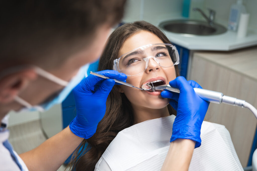
Endodontic intervention becomes necessary when bacterial infiltration compromises the dental pulp beyond reversible inflammation. Multiple clinical indicators manifest as the pulpal tissue undergoes necrosis and periapical tissues respond to bacterial toxins. Early recognition of these pathological signs determines treatment success rates and prevents systemic complications. Understanding the specific symptoms that differentiate reversible pulpitis from irreversible damage remains critical for timely intervention before tooth extraction becomes the only viable option.
Persistent or Severe Tooth Pain That Won’t Go Away
Persistent dental pain represents one of the most definitive indicators that endoscopic root canal therapy may be necessary. This symptomatology typically manifests as unrelenting discomfort localized to a specific tooth, often intensifying during mastication or upon application of thermal stimuli. Patients frequently report sharp shooting pains radiating along the trigeminal nerve pathways, indicating irreversible pulpitis or periapical pathology.
The pain’s etiology stems from bacterial infiltration of the pulp chamber, causing inflammation and subsequent necrosis of neurovascular tissues. Sudden onset pain occurring without provocation suggests acute pulpal degeneration requiring immediate intervention. Clinical examination reveals percussion sensitivity and potential periapical radiolucency on radiographic imaging. Unlike reversible pulpitis, which responds to conservative management, persistent pain lasting beyond 72 hours typically necessitates endodontic treatment to eliminate infected tissue and preserve tooth structure.
Prolonged Sensitivity to Hot and Cold Temperatures
Dental hypersensitivity that persists beyond the normal 5-10 second response indicates potential pulpal inflammation or necrosis requiring endodontic intervention. The affected tooth typically exhibits prolonged discomfort lasting 30 seconds or longer following exposure to thermal stimuli, distinguishing pathological sensitivity from routine dentinal hypersensitivity. Both cold and hot temperature triggers can elicit this response, though sensitivity to heat often correlates with more advanced pulpal degradation and irreversible pulpitis.
Duration of Sensitivity
When thermal sensitivity extends beyond the typical 1-2 second response following exposure to temperature stimuli, this prolonged reaction often indicates irreversible pulpitis or pulpal necrosis requiring endodontic intervention. Normal dental tissues exhibit brief thermal responses that dissipate immediately upon stimulus removal. Pathological pulpal conditions manifest as lingering discomfort persisting 30 seconds or longer after temperature exposure cessation.
Clinicians assess duration of pain through controlled thermal testing protocols. Patients experiencing intermittent sensitivity episodes lasting several minutes following hot or cold contact demonstrate compromised pulpal change. The transition from reversible to irreversible pulpitis occurs when inflammatory mediators overwhelm the pulp’s reparative capacity. Extended sensitivity duration correlates directly with bacterial infiltration depth and inflammatory cascade progression. Documentation of pain persistence patterns assists practitioners in differentiating between reversible conditions amenable to conservative treatment and irreversible pathology necessitating root canal therapy.
Types of Temperature Triggers
Although various thermal stimuli can elicit pulpal responses, specific temperature ranges and exposure sources provide diagnostic insight into endodontic pathology severity. Cold stimuli below 15°C typically trigger sharp, transient pain in reversible pulpitis, while temperatures exceeding 45°C produce dull, lingering discomfort indicative of critical damage. Common triggers include ice water, hot beverages, ambient air exposure, and dietary temperature variations encountered during mastication.
Clinicians evaluate pulpal essentiality through controlled temperature fluctuations using refrigerant spray (-26°C) or heated gutta-percha (65°C). Patients requiring endodontic intervention demonstrate hypersensitivity to both extremes, with pain persisting beyond stimulus removal. The intensity and duration of response correlate with inflammation degree and neural involvement. Differential diagnosis relies on distinguishing between normal dentin sensitivity and pathological pulpal reactions through systematic thermal testing protocols.
Tooth Discoloration or Darkening
Discoloration of dental enamel manifests as a progressive darkening that typically indicates pulpal necrosis or hemorrhage within the tooth structure. This visible tooth discoloration occurs when blood vessels rupture inside the pulp chamber, releasing hemoglobin that infiltrates dentinal tubules. The degradation of these blood products creates gray, brown, or black pigmentation.
Tooth appearance changes develop gradually following trauma or advanced decay. Internal resorption accelerates chromogenic transformation as necrotic tissue accumulates within the canal system. Clinicians differentiate intrinsic staining from external discoloration through transillumination and radiographic evaluation.
Periapical pathology correlates with color alterations in 92% of cases requiring endodontic intervention. Early detection prevents abscess formation and preserves remaining viable tissue. Spectrophotometric analysis confirms diagnosis when subtle changes escape visual examination.
Swollen or Tender Gums Near the Affected Tooth
Periapical inflammation extending from infected pulpal tissue causes localized gingival swelling and tenderness adjacent to compromised teeth. The bacterial toxins and inflammatory mediators migrate through the apical foramen, establishing periradicular pathosis that manifests as palpable gingival edema. Patients typically report discomfort upon digital pressure application to the affected area.
The inflamed gum tissue presents with erythema, edema, and possible suppuration when infection progresses to abscess formation. Chronic periapical periodontitis may induce gum recession through sustained inflammatory processes destroying supporting periodontal structures. Clinical examination reveals vestibular swelling corresponding to the affected root apex, with percussion sensitivity confirming pulpal involvement.
These gingival manifestations indicate advanced endodontic pathology requiring immediate intervention. Radiographic assessment combined with thermal and electric pulp testing confirms diagnosis, while prompt root canal therapy prevents further periodontal deterioration and systemic complications.
A Recurring Pimple or Bump on Your Gums
A dental abscess may manifest as a persistent fistula, appearing as a small, pimple-like lesion on the gingival tissue adjacent to the infected tooth. This draining sinus tract typically presents as a raised, yellowish-white pustule measuring 2-5 millimeters in diameter with a central opening that intermittently discharges purulent exudate. The fistula forms when bacterial infection within the tooth’s pulp chamber or periapical region creates sufficient pressure to establish a drainage pathway through the alveolar bone and soft tissue to the oral cavity.
What It Looks Like
When examining the oral cavity for signs of endodontic pathology, clinicians and patients may observe a persistent fistulous tract manifesting as a small, raised lesion on the gingival tissue adjacent to the affected tooth. This periapical abscess presents as a yellowish-white pustule, typically 2-5 millimeters in diameter, resembling an oral pimple. The lesion exhibits distinct visual changes, including erythematous surrounding tissue and potential purulent discharge upon palpation.
The fistula’s location correlates directly with the compromised tooth’s root apex, indicating severe deterioration of the pulpal tissue’s structural integrity. These draining sinus tracts may fluctuate in size, occasionally rupturing to release accumulated exudate before reforming. The gingival surface surrounding the lesion often appears inflamed, displaying characteristic edema and tissue blanching when pressure is applied.
Why It Forms
The formation of these gingival lesions results from bacterial colonization within the necrotic pulpal chamber, triggering an inflammatory cascade that extends beyond the tooth’s apical foramen into surrounding periradicular tissues. Persistent tooth decay formation creates pathways for microorganisms to penetrate dentinal tubules, ultimately reaching the pulpal space. Once established, anaerobic bacteria proliferate within devitalized tissue, producing toxic metabolites and endotoxins.
These inflammation triggers activate neutrophils and macrophages, generating purulent exudate that accumulates at the root apex. Hydrostatic pressure forces this suppurative material through the path of least resistance, typically perforating the buccal cortical plate. The resulting fistulous tract terminates at the gingival surface, manifesting as a draining sinus. Chronic periapical periodontitis perpetuates this process, with intermittent drainage preventing acute abscess formation while maintaining the pathological communication between infected root and oral cavity.
Pain When Chewing or Applying Pressure
Localized discomfort during mastication or upon application of occlusal force represents a cardinal indicator of potential endodontic pathology. This symptomatology typically manifests when bacterial infiltration compromises the periapical tissues surrounding the affected tooth’s root apex.
The mechanoreceptors within the periodontal ligament become hypersensitive to compression forces, triggering nociceptive responses even during routine masticatory function. Patients frequently report increased sensitivity when consuming harder foodstuffs or inadvertently clenching. The inflammation extends beyond the tooth structure, often producing a tender jaw sensation in the adjacent musculature and osseous tissues.
Clinical examination reveals percussion sensitivity and palpation tenderness over the root apex. The pain characteristically intensifies with vertical pressure rather than lateral forces, distinguishing it from periodontal pathology. Without intervention, this mechanical allodynia progressively worsens, ultimately rendering the tooth non-functional for mastication.
Cracked or Chipped Tooth With Exposed Nerves
Structural compromise of dental enamel and dentin through fracture or fragmentation frequently creates direct pathways to the pulpal chamber, exposing neurovascular tissues to external stimuli. Such tooth structure damage necessitates immediate clinical evaluation to prevent bacterial infiltration and subsequent pulpal necrosis.
Traumatic incidents resulting in crown fractures often reveal exposure of inner tooth layers, particularly when fracture lines extend below the gingival margin. Patients typically present with acute thermal sensitivity, spontaneous pain, and visible pulpal tissue. Microfractures, though less apparent during visual examination, permit bacterial penetration through dentinal tubules, initiating inflammatory cascades within the pulp.
Diagnostic radiographs and transillumination assist in identifying fracture depth and pulpal involvement. Without timely endodontic intervention, exposed dental pulp undergoes irreversible degeneration, necessitating root canal therapy to preserve tooth functionality and prevent periapical pathology.
Previous Deep Fillings or Dental Work Complications
Restorative procedures extending into deep cavity preparations near the pulpal floor frequently precipitate delayed endodontic complications, particularly when remaining dentin thickness measures less than 0.5 millimeters. Pre existing restorations compromise pulpal essential vitality through mechanical trauma, thermal conductivity changes, and microleakage pathways facilitating bacterial infiltration.
Dental history complications manifest as progressive pulpal degeneration following extensive amalgam or composite placements. Clinical indicators include persistent sensitivity, spontaneous pain, and periapical radiolucencies developing months to years post-treatment. Large restorations alter stress distribution patterns, creating microfractures that propagate toward pulpal tissues.
Secondary caries beneath existing restorations necessitate deeper excavation, often breaching pulpal boundaries. Recurrent operative interventions generate cumulative trauma, depleting reparative dentin formation capacity. Histopathological examination reveals chronic inflammatory infiltrates, dystrophic calcification, and eventual pulpal necrosis requiring endodontic intervention.
Frequently Asked Questions
How Much Does a Root Canal Typically Cost With and Without Insurance?
The average cost of root canal therapy ranges from $700-$1,500 without insurance, depending on tooth location. Dental insurance coverage for root canals typically reimburses 50-80% of procedural fees, reducing patient responsibility to $200-$750.
How Long Does a Root Canal Procedure Take to Complete?
A root canal procedure typically requires 60-90 minutes per appointment. Complex cases necessitate multiple visits. Recovery timeline after root canal involves 2-4 days. Patients should monitor for symptoms of root canal complications including persistent pain or swelling.
What Happens if I Delay or Avoid Getting a Needed Root Canal?
Delaying necessary endodontic treatment results in progressive pulpal necrosis with increased risk of infection, periapical abscess formation, and systemic complications. Untreated cases frequently culminate in potential tooth loss, necessitating extraction and subsequent prosthetic rehabilitation.
Are There Any Alternatives to Root Canal Treatment?
Alternative tooth preservation options include pulp capping procedures and pulpotomy as minimally invasive treatments for reversible pulpitis. However, extraction with subsequent implant placement remains the definitive alternative when endodontic therapy proves unsuitable for irreversible pulpal pathology.
How Painful Is a Root Canal Compared to Tooth Extraction?
Root canal therapy typically produces less intraoperative discomfort than extraction procedures. Recovery time after procedure favors endodontic treatment, with minimal postoperative sequelae. Extraction sites demonstrate prolonged healing periods, while root canal patients experience reduced level of discomfort.