
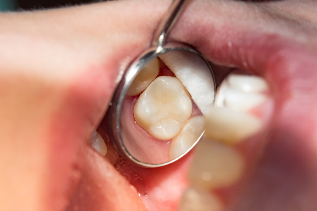
Dental restorations become necessary when carious lesions penetrate the enamel layer and compromise dentin integrity. Clinical indicators include persistent pulpal sensitivity, visible cavitation, and radiographic evidence of demineralization extending beyond the dentinoenamel junction. Without intervention, bacterial infiltration accelerates structural degradation, potentially requiring endodontic treatment or extraction. The critical threshold for restoration depends on lesion depth, patient caries risk, and progression rate—factors that determine whether conservative management remains viable.
Common Signs and Symptoms That Indicate You Need a Filling
When tooth structure becomes compromised by bacterial decay or physical damage, several clinical indicators typically manifest that suggest the need for restorative intervention. Persistent toothache symptoms represent the most common presenting complaint, ranging from sharp, localized pain during mastication to spontaneous throbbing sensations indicating pulpal involvement. Visible tooth discoloration, particularly brown or black spots on occlusal surfaces or interproximal regions, signifies active carious lesions requiring immediate attention.
Additional diagnostic markers include heightened sensitivity to thermal stimuli, food impaction in cavitated areas, and palpable surface irregularities detected during routine examination. Patients may report discomfort when consuming sweet substances, indicating exposed dentin tubules. Radiographic evidence often reveals radiolucent areas beneath enamel surfaces before clinical symptoms emerge. Early intervention prevents progression to more extensive restorative procedures or endodontic treatment.
How Dentists Detect and Diagnose Cavities During Examinations
Although patients may present with symptomatic complaints, thorough cavity detection requires systematic clinical evaluation employing multiple diagnostic modalities. Dentists initiate examinations through visual inspection, identifying discoloration, structural defects, and plaque buildup detection along tooth surfaces. Tactile exploration using sharp explorers reveals sticky areas indicating demineralization. Radiographic imaging captures interproximal and subgingival caries undetectable through visual means.
Advanced diagnostic technologies enhance detection accuracy. Digital radiography provides enhanced resolution while minimizing radiation exposure. Laser fluorescence devices quantify bacterial byproducts within lesions. Transillumination identifies enamel microfractures and early-stage decay. During extensive examinations, practitioners perform gum health assessment to evaluate periodontal conditions affecting restorative treatment planning. Clinical findings are systematically documented, establishing baseline measurements for monitoring disease progression and determining intervention timing.
Different Types of Tooth Damage That Require Fillings
Dental fillings address multiple forms of structural tooth damage beyond standard carious lesions. Cavities resulting from bacterial demineralization, fractured tooth surfaces from occlusal trauma or thermal stress, and abraded enamel regions from mechanical wear all necessitate restorative intervention. Each pathological condition requires specific clinical assessment to determine the appropriate filling material and restoration technique.
Cavities and Decay
Most dental fillings address carious lesions that manifest through progressive demineralization of tooth enamel and subsequent dentin involvement. Bacterial acid production initiates enamel erosion, creating microscopic pores that expand into visible cavitations. Early-stage caries presents as chalky white spots, while advanced lesions exhibit brown or black tooth discoloration indicating deeper bacterial penetration.
Dentists diagnose decay through visual examination, radiographic analysis, and tactile exploration using specialized instruments. Untreated caries progresses through three distinct stages: enamel demineralization, dentin invasion, and pulpal exposure. While unusual growths rarely occur within carious lesions, secondary complications include periapical abscesses and cyst formation. Restorative intervention becomes essential when cavitation breaches the dentinoenamel junction. The extent of decay determines filling material selection, with composite resins preferred for minor lesions and amalgam or ceramic inlays recommended for extensive structural compromise.
Cracked Tooth Surfaces
Why do seemingly minor tooth fractures necessitate immediate restorative treatment? Cracked tooth surfaces compromise the structural integrity of the tooth, creating pathways for bacterial infiltration into deeper dental tissues. These microfractures often originate from occlusal trauma, thermal stress, or parafunctional habits such as bruxism.
Fractured tooth enamel exposes underlying dentin, increasing sensitivity and accelerating deterioration. Without intervention, crack propagation extends through the enamel-dentin junction, potentially reaching the pulp chamber. Clinical examination reveals various fracture patterns: craze lines, fractured cusps, and vertical root fractures, each requiring specific restorative approaches.
Dental fillings effectively seal cracked tooth surfaces, preventing further damage while restoring anatomical form and function. Composite resins or amalgam materials bond to remaining tooth structure, distributing masticatory forces and halting crack progression before irreversible pulpal involvement occurs.
Worn Enamel Areas
How does enamel erosion progress from microscopic surface changes to significant structural loss requiring restorative intervention? Initial demineralization occurs at the molecular level when acids dissolve hydroxyapatite crystals, creating micropores in the enamel matrix. Progressive acid exposure leads to enamel thinning, manifesting as translucency at incisal edges and surface roughening. Exposed enamel becomes increasingly susceptible to mechanical wear from mastication and chemical dissolution from dietary acids.
Clinical evaluation reveals characteristic patterns: cupping lesions on occlusal surfaces, cervical notching, and generalized surface texture alterations. Advanced erosion exposes underlying dentin, presenting as yellow discoloration and thermal sensitivity. Restorative intervention becomes necessary when enamel loss exceeds 0.5mm depth, compromising structural integrity. Composite resin fillings restore anatomical contours, protect exposed dentin tubules, and prevent further deterioration of compromised tooth structure.
The Stages of Tooth Decay and When Fillings Become Necessary
When enamel demineralization exceeds the rate of remineralization, tooth decay progresses through distinct pathological stages that determine the necessity and timing of restorative intervention. Initial carious lesions manifest as white spot formations, representing subsurface porosity without cavitation. These early warning signs indicate reversible demineralization treatable through remineralization protocols.
Progressive bacterial acid production advances decay into dentin, creating irreversible structural damage requiring operative treatment. The caries penetration depth determines restoration necessity: enamel-limited lesions may arrest with fluoride therapy, while dentinal involvement mandates excavation and filling placement. Underlying issues such as xerostomia, dietary factors, or compromised oral hygiene accelerate progression rates. Clinical assessment utilizing radiographic examination and visual-tactile evaluation establishes decay severity. Intervention timing remains critical—premature restoration damages healthy tissue, while delayed treatment risks pulpal involvement and extensive tooth structure loss.
What Happens If You Delay Getting a Needed Filling
Untreated dental caries progress through dentin at an accelerated rate, expanding both laterally and toward the pulp chamber as bacterial acids continue demineralizing tooth structure. The enlarging cavity creates microchannels that expose dentinal tubules to thermal and chemical stimuli, resulting in sharp pain responses and persistent sensitivity to temperature variations. Without intervention, the carious lesion compromises structural integrity while approaching the neurovascular bundle, increasing the probability of irreversible pulpitis and subsequent endodontic treatment.
Cavity Growth Progression
What begins as microscopic demineralization rapidly accelerates into irreversible tooth destruction when dental caries remain untreated. Initial enamel porosity expands through bacterial acid production, creating subsurface lesions that advance toward dentin. The cavity progression stages follow predictable patterns: white spot lesions transform into cavitated defects, penetrating through enamel’s crystalline structure within months.
Once bacteria breach the dentinoenamel junction, cavity progression severity intensifies exponentially. Dentinal tubules provide pathways for rapid bacterial infiltration, accelerating destruction rates threefold compared to enamel degradation. Untreated lesions reaching pulpal proximity trigger inflammatory responses, potentially causing irreversible pulpitis within six to twelve months.
Advanced caries compromise structural integrity, increasing fracture susceptibility and necessitating extensive restorative interventions. Delayed treatment transforms manageable lesions requiring simple restorations into complex cases demanding endodontic therapy or extraction.
Pain and Sensitivity
Why does dental sensitivity progressively worsen following caries penetration through protective enamel barriers? Once bacterial invasion compromises dentin tubules, exposed nerve endings within the pulp chamber respond aggressively to external stimuli. Initial manifestations present as sudden temperature sensitivity when consuming hot or cold substances, indicating dentin exposure and tubular fluid movement triggering mechanoreceptor activation.
As demineralization advances toward the pulp, patients experience chronic aching pain resulting from inflammatory mediator release and increased intrapulpal pressure. Bacterial toxins stimulate nociceptors, producing spontaneous pain episodes lasting minutes to hours. Without intervention, reversible pulpitis progresses to irreversible inflammation, characterized by lingering discomfort after stimulus removal. Nocturnal pain intensifies due to positional blood flow changes. Advanced decay approaching the pulp horn triggers severe, throbbing pain requiring immediate endodontic assessment beyond simple restorative procedures.
Modern Filling Materials and Your Treatment Options
Today’s restorative dentistry offers multiple filling materials, each with distinct physical properties, clinical indications, and longevity profiles that influence treatment selection. Composite resins demonstrate superior adhesive qualities through chemical bonding to tooth structure, providing excellent marginal seal and minimal microleakage. These tooth-colored biocompatible materials polymerize under specific wavelength light, achieving immediate functional restoration.
Amalgam fillings, containing silver, tin, copper, and mercury alloy, deliver exceptional durability for posterior restorations subjected to high occlusal forces. Glass ionomer cements release fluoride ions, offering cariostatic properties ideal for pediatric applications and root surface lesions. Ceramic inlays and onlays, fabricated through CAD/CAM technology or laboratory processes, provide ideal aesthetics and wear resistance. Material selection depends on cavity size, location, patient allergies, aesthetic requirements, and cost considerations, requiring thorough clinical evaluation.
The Filling Procedure: What to Expect During Your Appointment
How dental professionals approach cavity restoration follows established clinical protocols designed to safeguard ideal treatment outcomes and patient comfort. The procedure initiates with comprehensive radiographic assessment and visual examination to determine decay extent and treatment parameters.
The numbing process involves topical anesthetic application followed by local anesthetic injection, typically lidocaine with epinephrine, administered via inferior alveolar or infiltration techniques. Complete anesthesia verification precedes operative procedures.
Clinicians employ rubber dam isolation to maintain aseptic conditions while removing carious tissue using high-speed handpieces with carbide or diamond burs. Cavity preparation follows specific geometric principles ensuring adequate retention and resistance form.
Pain management extends beyond initial anesthesia through careful tissue manipulation and appropriate restoration technique. Material placement occurs incrementally, with photopolymerization or chemical curing completing the process. Final occlusal adjustment guarantees proper bite alignment.
Preventing Future Fillings Through Proper Oral Care
When implemented systematically, evidence-based preventive strategies substantially reduce caries incidence and subsequent restorative requirements. Effective dental hygiene habits constitute the primary defense mechanism against bacterial biofilm accumulation and demineralization processes. Patients should execute thorough brushing twice daily with fluoridated dentifrice, employing modified Bass technique for ideal plaque disruption. Interproximal cleaning via floss or interdental brushes removes debris from areas inaccessible to conventional brushing.
Regular dental check ups facilitate early detection of incipient lesions before cavitation necessitates operative intervention. Professional prophylaxis eliminates calcified deposits and provides fluoride varnish application when indicated. Dietary modification, particularly limiting fermentable carbohydrate frequency, disrupts acidogenic bacterial metabolism. Prescription-strength fluoride supplements or xylitol products may benefit high-risk individuals. These combined protocols demonstrate significant efficacy in preventing progression from reversible demineralization to irreversible tooth structure loss requiring restoration.
Frequently Asked Questions
How Much Does a Dental Filling Typically Cost?
Dental filling costs range from $50-$450 per tooth, determined by filling material options including amalgam, composite resin, or ceramic. Dental insurance coverage typically reimburses 50-80% of procedural fees after deductible requirements are satisfied by patients.
Does Getting a Filling Hurt?
Patients typically experience minimal pain due to local anesthetic administration creating a numbing sensation during procedure. Post-operative symptoms include temporary discomfort after procedure, manageable with over-the-counter analgesics. Modern techniques guarantee the filling process remains relatively painless.
How Long Do Dental Fillings Usually Last?
Dental filling longevity varies considerably: amalgam restorations typically endure 10-15 years, while composite materials last 5-10 years. Proper dental hygiene, including biannual prophylaxis and ideal homecare protocols, substantially enhances restoration durability and prevents secondary caries formation.
Can I Eat Immediately After Getting a Filling?
Patients should wait 2-24 hours before eating, depending on filling material. Immediate consumption risks compromising restoration integrity. Post-operative sensitivity to hot/cold temperatures and difficulty chewing on filled tooth typically resolve within 48-72 hours following procedure.
Will My Dental Insurance Cover the Cost of Fillings?
Most dental insurance plans cover 50-80% of amalgam or composite filling procedures after deductibles. Patients typically encounter out-of-pocket costs for fillings due to dental insurance coverage limitations, annual maximums, and material upgrade fees for tooth-colored restorations.