A routine dental examination follows a systematic protocol designed to assess oral health thoroughly. The procedure begins with administrative tasks and medical history documentation before progressing to clinical interventions. Dental professionals employ specialized instruments and diagnostic technologies to evaluate both hard and soft tissues within the oral cavity. Each component of the examination serves a specific diagnostic purpose, from detecting subclinical pathology to identifying periodontal disease indicators that may require immediate intervention.
The Check-In Process and Medical History Review
Upon arrival at the dental office, patients complete essential administrative tasks that establish the foundation for their clinical care. Reception personnel verify insurance eligibility, update demographic information, and collect copayments. Patients receive thorough medical history questionnaires documenting systemic conditions, current medications, allergies, and previous dental treatments. Privacy concerns are addressed through HIPAA compliance protocols, ensuring confidential handling of protected health information.
Communication with staff includes disclosure of chief complaints, symptom duration, and pain characteristics. Clinical personnel review cardiovascular conditions, bleeding disorders, and immunosuppressive states that may necessitate prophylactic antibiotics or treatment modifications. Pregnancy status, bisphosphonate therapy, and recent hospitalizations require documentation. Staff members clarify medication interactions, particularly anticoagulants affecting surgical procedures. This systematic data collection enables risk stratification and appropriate treatment planning before clinical examination begins.
Professional Teeth Cleaning and Plaque Removal
Professional teeth cleaning constitutes a critical component of the dental examination, employing specialized instruments to eliminate calcified deposits and bacterial biofilm from tooth surfaces. The hygienist performs scaling to remove supragingival and subgingival tartar using ultrasonic scalers or hand instruments, followed by polishing with a rotary handpiece and prophylactic paste to eliminate residual plaque and surface stains. The procedure concludes with interproximal flossing to remove debris from contact points and assess gingival response to mechanical stimulation.
Scaling and Tartar Removal
When calcified deposits accumulate on tooth surfaces despite routine oral hygiene measures, dental professionals perform scaling procedures to mechanically remove these mineralized biofilms. Hygienists utilize ultrasonic scalers operating at 25,000-30,000 Hz frequencies, generating cavitation bubbles that disrupt tartar adhesion while irrigating debris. Manual instrumentation follows, employing area-specific curettes with 70-80 degree cutting edges to access subgingival deposits.
Removal techniques vary based on deposit thickness and location. Supragingival scaling addresses visible calculus using sickle scalers and universal curettes. Subgingival procedures require periodontal probes to measure pocket depths before systematic root surface cleansing. Cross-hatching strokes guarantee complete calculus elimination from proximal surfaces and furcation areas. Post-scaling polishing with prophylaxis paste removes residual stains while smoothing microscopic surface irregularities. This thorough approach disrupts bacterial colonization patterns, supporting effective plaque buildup prevention between appointments.
Polishing Your Teeth
Following calculus removal, dental professionals implement polishing protocols to eliminate residual plaque biofilm and extrinsic staining from enamel surfaces. The hygienist selects appropriate prophylaxis paste containing specific abrasive particles based on patient-specific staining severity and enamel condition. Rubber cup attachments or air-polishing devices deliver controlled mechanical action across tooth surfaces.
Standard polishing technique involves systematic progression through dental quadrants, applying intermittent pressure at 2,500-3,000 RPM rotational speeds. Clinicians maintain ideal angulation while addressing all accessible surfaces—buccal, lingual, and occlusal. Typical polishing duration ranges from 2-5 minutes per arch, depending on stain accumulation and surface irregularities. The procedure concludes with thorough irrigation to remove paste residue and debris. This final step enhances surface smoothness, reduces bacterial adhesion potential, and provides temporary fluoride uptake when fluoridated pastes are utilized.
Flossing Between Teeth
After completing surface polishing, dental hygienists initiate interproximal cleaning procedures using specialized flossing techniques to access biofilm deposits between adjacent teeth. Professional interproximal debridement removes bacterial colonies and calcified deposits unreachable through conventional brushing protocols.
Hygienists select appropriate dental floss options based on interdental spacing, restoration presence, and tissue sensitivity. Waxed floss facilitates navigation through tight contact points, while unwaxed variants provide superior plaque disruption. Specialized alternatives include polytetrafluoroethylene tape for wide embrasures and superfloss for fixed prosthetics.
Proper flossing technique involves C-shaped contouring against proximal tooth surfaces, applying controlled apical-coronal strokes beneath gingival margins. Hygienists systematically advance through each interproximal space, maintaining tension while avoiding traumatic snapping motions. This methodical approach guarantees thorough biofilm elimination from mesial and distal surfaces, preventing periodontal pathology development in these high-risk zones.
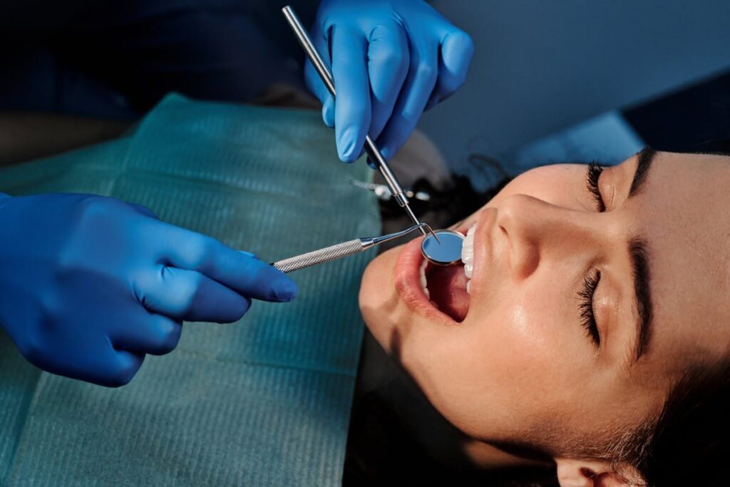
Comprehensive Oral Examination by Your Dentist
Following the prophylaxis procedure, the dentist conducts a systematic visual examination of all tooth surfaces using a dental explorer and mouth mirror to identify caries, fractures, and existing restorations. The practitioner evaluates periodontal tissues through visual inspection and periodontal probing to measure pocket depths, assess gingival recession, and detect signs of inflammation or bleeding. This clinical assessment establishes baseline measurements for monitoring oral health status and determining necessary interventions.
Visual Tooth Assessment
The dentist initiates the visual tooth assessment by systematically examining each tooth surface using a dental mirror and explorer instrument. This methodical evaluation identifies caries, fractures, and structural defects across all dentition. The practitioner observes tooth discoloration patterns that may indicate decay, previous restorations, or intrinsic staining requiring intervention.
During examination, the dentist evaluates enamel condition by detecting surface irregularities, demineralization zones, and erosion patterns. The explorer instrument detects tactile changes in tooth texture, including soft spots suggesting active caries or rough areas indicating early lesions. Each tooth receives individual documentation using standardized charting systems, recording existing restorations, abnormalities, and treatment needs. The assessment includes occlusal, buccal, lingual, mesial, and distal surfaces, ensuring thorough evaluation of the entire dental arch for accurate diagnosis and treatment planning.
Gum Health Check
Beyond tooth surface evaluation, periodontal assessment constitutes a critical component of thorough oral examination. The dentist employs a periodontal probe to measure pocket depths between teeth and gingival tissue, recording measurements at six points per tooth. Normal sulcus depth ranges from 1-3 millimeters; readings exceeding this threshold indicate potential periodontal disease.
During examination, the practitioner evaluates tissue color, texture, and consistency. Healthy gingiva appears pink and firm, while gum inflammation manifests as redness, swelling, or bleeding upon probing. The clinician documents areas of gum recession, measuring attachment loss from the cementoenamel junction. Additional assessment includes checking for tooth mobility, furcation involvement, and suppuration. Radiographs complement clinical findings by revealing bone levels and calculus deposits. This systematic evaluation enables early detection of periodontal pathology and appropriate treatment planning.
Digital X-Rays and Diagnostic Imaging
Digital radiographic technology has revolutionized oral diagnostic capabilities by providing immediate, high-resolution images with substantially reduced radiation exposure compared to traditional film-based systems. Bitewing radiographs capture interproximal surfaces between adjacent teeth, enabling detection of proximal caries, calculus deposits, and crestal bone levels. These intraoral projections typically encompass premolar and molar regions bilaterally.
Panoramic imaging generates all-encompassing extraoral views displaying maxillary and mandibular arches, temporomandibular joints, and surrounding anatomical structures within a single exposure. Periapical radiographs visualize complete tooth structure including apex and periapical tissues for endodontic assessment. Digital sensors eliminate chemical processing, reduce retake frequency, and facilitate image enhancement through contrast adjustment and magnification. Diagnostic imaging frequency depends on individual risk factors, clinical findings, and American Dental Association guidelines, ranging from six-month intervals for high-risk patients to 24-36 months for asymptomatic adults.
Oral Cancer Screening and Soft Tissue Assessment
While radiographic evaluation provides critical subsurface information, exhaustive examination requires systematic visual and tactile assessment of oral mucosa, identifying potentially malignant lesions during earliest developmental stages. The practitioner employs bimanual palpation techniques, evaluating lymph nodes, salivary glands, and floor of mouth for asymmetry, induration, or masses.
Tongue inspection involves dorsal, ventral, and lateral border examination using gauze retraction, noting color variations, texture abnormalities, or suspicious ulcerations persisting beyond two weeks. The clinician performs thorough throat examination, visualizing tonsillar pillars, posterior pharyngeal wall, and soft palate while documenting erythematous patches, leukoplakia, or erythroplakia presentations.
Additional screening encompasses labial mucosa, buccal vestibules, and gingival tissues. Adjunctive technologies including fluorescence visualization or brush biopsy may supplement conventional examination when atypical findings warrant enhanced diagnostic precision.
Gum Health Evaluation and Periodontal Measurements
Following exhaustive soft tissue assessment, the examiner initiates periodontal evaluation through systematic probing of gingival sulci using a calibrated periodontal probe, measuring pocket depths at six sites per tooth while recording bleeding points, suppuration, and recession levels. The clinician performs thorough gingival inflammation assessment, documenting tissue color, texture, and contour deviations from physiologic norms. Probing pocket depths exceeding three millimeters indicate potential periodontal pathology requiring intervention.
The practitioner evaluates clinical attachment levels, calculating differences between cemento-enamel junction and sulcus base measurements. Furcation involvement receives classification grades, while mobility testing determines tooth stability through standardized force application. Radiographic analysis supplements clinical findings, revealing osseous defects and alveolar bone loss patterns. Documentation includes plaque indices, bleeding scores, and recession measurements, establishing baseline parameters for disease progression monitoring and treatment planning protocols.
Discussion of Findings and Treatment Recommendations
Upon completion of detailed oral examination and data collection, the dentist synthesizes clinical findings into a coherent diagnostic summary presented to the patient using clear anatomical references and visual aids. Radiographic images, intraoral photographs, and periodontal charts facilitate extensive understanding of existing pathology and structural concerns.
Treatment planning protocols prioritize interventions based on clinical urgency, establishing immediate, short-term, and long-term therapeutic objectives. The dentist delineates restorative options, including direct composite restorations, indirect ceramic reconstructions, endodontic therapy, periodontal interventions, and prosthetic rehabilitation when indicated.
Cost considerations integrate insurance coverage parameters, out-of-pocket expenses, and phased treatment alternatives. Financial coordinators provide detailed fee schedules, pre-authorization requirements, and payment structuring options. Informed consent documentation encompasses procedural risks, benefits, alternative treatments, and prognosis without intervention, ensuring patient autonomy in clinical decision-making processes.
Scheduling Follow-Up Care and Next Appointments
After establishing thorough treatment protocols, the dental practice implements systematic appointment scheduling based on clinical priorities and periodontal maintenance intervals. The administrative staff coordinates next visit planning according to diagnosed pathology severity, insurance authorization timelines, and procedural sequencing requirements. Urgent restorative needs receive immediate scheduling priority, while preventive maintenance appointments follow standard six-month recall protocols unless periodontal conditions necessitate quarterly evaluations.
The scheduling coordinator documents specific procedural codes, estimated treatment duration, and pre-appointment requirements in the practice management system. Patients receive written appointment confirmations detailing scheduled procedures, financial obligations, and pre-treatment instructions. The office establishes automated reminder protocols through digital communication platforms to minimize appointment failures. Follow-up scheduling integrates with the treatment plan timeline, ensuring continuity of care while accommodating practice capacity and specialist referral coordination when indicated.
Frequently Asked Questions
How Much Does a Routine Dental Exam Typically Cost Without Insurance?
The average cost range for routine dental examinations without insurance spans $50-$350, depending on multiple cost factors including geographic location, practitioner credentials, diagnostic imaging requirements, and detailed periodontal assessment protocols included within the examination scope.
Can I Eat or Drink Before My Dental Exam Appointment?
Patients typically require no fasting requirements before routine dental examinations. Hydration recommendations include consuming water freely. Clinicians advise avoiding food particles immediately prior, though light meals remain acceptable. Specific procedural restrictions apply only for sedation-based interventions.
How Often Should I Schedule Routine Dental Exams?
Patients with ideal dental hygiene habits should schedule biannual examinations. Dental check up frequency increases to quarterly intervals for individuals with periodontal disease, extensive restorative work, or compromised oral health requiring enhanced clinical surveillance and intervention.
What Should I Do if I Have Dental Anxiety?
Patients experiencing dental anxiety should practice relaxation techniques including controlled breathing and progressive muscle relaxation. Scheduling a pre-appointment consultation allows discussion of sedation options, establishing signals, and familiarizing oneself with clinical procedures before actual treatment commences.
Can I Bring My Child With Me to My Appointment?
Patients may bring children to appointments; however, clinical protocols require ensuring child’s comfort through designated waiting areas. Bringing a child necessitates pre-appointment notification for appropriate accommodations, supervision arrangements, and compliance with practice-specific pediatric presence policies.