Dental X-Rays St, Graham, TX

Dental X-rays in Graham, TX, are a pivotal component of thorough oral care, offering dentists critical insights into a patient’s dental health. Utilizing state-of-the-art imaging technology, these X-rays assist in the early detection of dental issues, such as cavities and periodontal disease, ensuring timely and effective treatment. With strict adherence to safety protocols, patients are assured minimal radiation exposure. Discover how these essential diagnostic tools enhance dental health and what to expect during your appointment.
Understanding Dental X-Rays
Dental radiography, a vital diagnostic tool in modern dentistry, provides detailed imagery of oral structures that are invisible to the naked eye. Despite its essential role, several dental x-ray myths persist, causing unnecessary patient concerns. One common misconception is the belief that dental x-rays expose patients to high levels of radiation. In reality, advancements in technology have minimized exposure, making it comparable to natural environmental radiation levels. Another myth suggests that x-rays are only necessary for patients with symptoms, overlooking their preventive capabilities in detecting early-stage dental issues. Addressing these myths is significant for informed patient decision-making. Dentists are tasked with educating patients on the safety and necessity of dental radiography, ensuring that concerns are addressed with evidence-based information.
Types of Dental X-Rays
Dental radiography encompasses various types of X-rays, each serving specific diagnostic purposes. Bitewing X-rays are primarily utilized to detect interproximal caries and monitor restorations, while panoramic X-rays offer a thorough view of the entire oral cavity structure, assisting in evaluating jaw relationships and detecting abnormalities. Additionally, periapical X-rays provide detailed images of individual teeth and surrounding bone, essential for evaluating root and periodontal conditions.
Bitewing X-Rays Explained
While often overlooked in routine dental visits, bitewing X-rays serve as an indispensable diagnostic tool in preventive dentistry. These radiographs allow for the detailed examination of the interproximal areas of the posterior teeth, where decay and periodontal disease often originate. Proper bitewing placement is essential to accurately capture the crowns and upper parts of both the upper and lower teeth in a single image, ensuring peak diagnostic capability. The bitewing frequency is typically determined by the patient’s individual risk assessment, dental history, and clinical examination. Regular bitewing X-rays facilitate early detection of cavities, bone loss, and other dental pathologies, enabling timely intervention. This proactive approach is critical in maintaining oral health and preventing more extensive and costly dental treatments.
Panoramic X-Rays Benefits
How do panoramic X-rays enhance the depth of dental diagnostics? Panoramic X-rays offer a considerable advantage by providing a wide-ranging view of the entire oral cavity, including the teeth, jaws, and surrounding bone structures. This imaging technique captures a detailed dental assessment in a single scan, greatly reducing the necessity for multiple radiographs. Such efficiency is particularly beneficial for evaluating dental anomalies, impacted teeth, and jaw disorders with precision. By delivering high-resolution images, panoramic X-rays facilitate early detection of pathologies, thereby enhancing treatment planning and patient outcomes. Furthermore, this method is less invasive, minimizing patient discomfort while ensuring accurate diagnostics. Overall, the panoramic x ray advantages contribute to a more thorough and effective approach to dental care.
Periapical X-Rays Uses
Among the various types of dental X-rays, periapical X-rays hold a significant role in providing detailed insights into individual teeth and their surrounding bone structures. Periapical imaging is indispensable for capturing the entire tooth, from crown to root, thereby ensuring thorough examination. This type of imaging is particularly beneficial in diagnosing conditions such as dental abscesses, cysts, or bone loss. It aids in evaluating the health of the tooth’s root and surrounding bone, facilitating early detection of pathologies. By offering high diagnostic clarity, periapical X-rays enable clinicians to make informed decisions regarding endodontic treatment and surgical planning. Their precision supports accurate monitoring of treatment outcomes, ensuring ideal patient care through targeted intervention and improved prognostic evaluation.
The Process of Taking Dental X-Rays
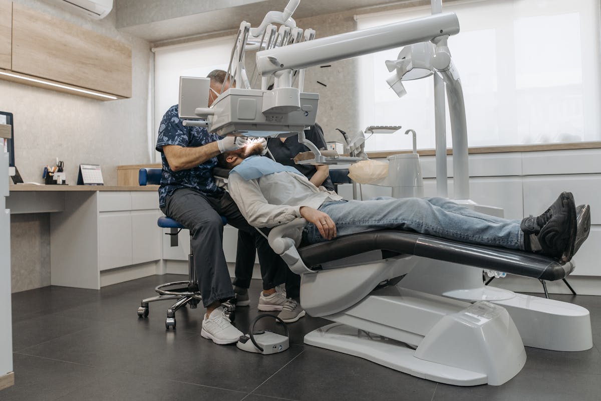
The process of taking dental X-rays involves careful positioning of the patient and the X-ray machine to guarantee accurate imaging. Dental professionals employ various X-ray techniques, such as bitewing and panoramic, to capture thorough views of the oral cavity. Proper alignment of the X-ray beam is essential to minimize distortion and enhance diagnostic accuracy. Ensuring patient comfort is paramount; therefore, practitioners use positioning devices and lead aprons to protect and stabilize patients during the procedure. The selection of appropriate technique depends on the specific diagnostic needs, balancing detailed imaging with reduced radiation exposure. Digital sensors or traditional film are positioned intraorally or extraorally, depending on the technique employed. The procedure is quick, minimizing discomfort while maximizing diagnostic value for the patient.
Benefits of Dental X-Rays
Dental X-rays serve as an indispensable diagnostic tool in modern dentistry, providing detailed insights into oral health that are otherwise invisible to the naked eye. These radiographs enable clinicians to identify and monitor conditions such as caries, periodontal disease, and root infections at an early stage, thereby facilitating timely intervention. The importance of awareness regarding the role of X-rays cannot be overstated, as they uncover hidden dental issues that could escalate without detection. Despite common X-ray misconceptions, these images offer a non-invasive means to assess bone density, detect impacted teeth, and evaluate jaw conditions. By accurately diagnosing underlying problems, dental X-rays enhance treatment planning, ensuring ideal patient outcomes and preserving oral health. The precision afforded by X-rays is unparalleled in dental diagnostics.
Safety Measures in Dental X-Rays
In the context of dental X-rays, minimizing radiation exposure is paramount to patient safety. This is achieved through the implementation of advanced imaging technology and adherence to the ALARA (As Low As Reasonably Achievable) principle. Additionally, the use of protective equipment such as lead aprons and thyroid collars further mitigates risk, ensuring patient well-being during diagnostic procedures.
Minimizing Radiation Exposure
Maximizing patient safety during dental X-rays necessitates the implementation of rigorous radiation protection protocols. Key strategies in minimizing radiation exposure involve adhering to the ALARA (As Low As Reasonably Achievable) principle, which emphasizes radiation safety and exposure reduction. Dental practitioners employ techniques such as collimation to limit the X-ray beam size, thereby reducing scatter radiation. Utilizing digital radiography over conventional methods further decreases radiation doses due to its enhanced sensitivity. Additionally, limiting the frequency of X-ray examinations based on individual patient needs is crucial in minimizing unnecessary exposure. Meticulous calibration and maintenance of X-ray equipment guarantee peak performance, hence reducing patient dose. These measures collectively contribute to the overarching goal of safeguarding patient health while achieving diagnostic efficacy.
Protective Equipment Use
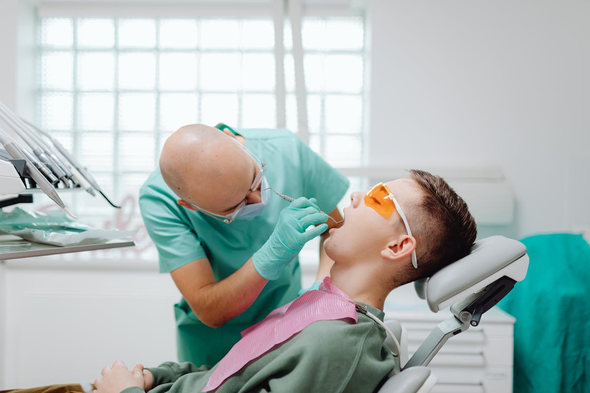
While ensuring patient safety during dental X-rays, the use of protective equipment plays a pivotal role. Protective barriers, such as lead aprons and thyroid collars, are essential components in minimizing radiation exposure. These barriers are specifically designed to shield vulnerable body areas, reducing the risk of radiation-induced damage. Dental practitioners adhere strictly to established safety protocols, ensuring each patient receives prime protection. The deployment of these barriers is a mandatory practice, aligned with regulatory standards and guidelines. In addition, advancements in protective technology further enhance their efficacy, providing robust defense mechanisms during radiographic procedures. By integrating these protective measures, dental professionals in Graham, TX, maintain a high standard of care, prioritizing patient well-being while executing essential diagnostic processes.
Identifying Dental Issues With X-Rays
Dental X-rays serve as an indispensable diagnostic tool, offering dental professionals a detailed view of oral structures that are otherwise hidden from the naked eye. Through meticulous x-ray interpretation, practitioners can identify a range of dental issues, including cavities, periodontal disease, and bone abnormalities. This diagnostic imaging technique plays an essential role in dental diagnosis by revealing the exact location and extent of these conditions, thereby guiding targeted treatment plans. Additionally, X-rays can detect impacted teeth, abscesses, and even early signs of oral cancer, ensuring proactive management of potential health threats. By providing an extensive visualization of the patient’s oral cavity, dental X-rays enhance the precision and effectiveness of dental care, ultimately contributing to improved patient outcomes and long-term oral health.
Technological Advancements in X-Ray Imaging
How have recent technological advancements transformed the landscape of dental X-ray imaging? The integration of digital imaging and sophisticated imaging software has greatly enhanced diagnostic accuracy. Advanced sensors now facilitate superior image quality while minimizing patient exposure through radiation reduction. These sensors, coupled with state-of-the-art image processing, allow for clearer visualization of dental structures, aiding precise diagnoses. Furthermore, the advent of 3D imaging has revolutionized imaging techniques, providing extensive views of the oral cavity that were previously unattainable. This three-dimensional perspective enables dentists to better assess complex conditions, improving treatment outcomes. Such advancements assure patients of a safer, more efficient diagnostic process, underscoring the importance of continually evolving technology in enhancing dental care practices.
Preparing for Your Dental X-Ray Appointment
As dental X-ray technology continues to advance, patients should be well-informed about how to prepare for their dental X-ray appointments to guarantee ideal results. Effective X-ray preparation tips include wearing comfortable clothing and removing any metallic objects, such as jewelry, that could interfere with image clarity. Additionally, patients should communicate any concerns or allergies to the dental technician. To manage patient anxiety, practitioners recommend arriving early to allow time for relaxation, understanding the procedure’s steps, and employing deep-breathing techniques. Familiarity with the process can alleviate apprehension, ensuring a smoother experience. It is essential for patients to follow instructions given by their dental provider, as this enhances the diagnostic accuracy of X-ray results and contributes to best oral health outcomes.
Choosing the Right Dental Practice in Graham, TX
When seeking ideal dental care in Graham, TX, what factors should patients consider to confirm they select the most suitable practice? Evaluating patient reviews provides critical insights into the experiences of others and the practice’s reputation. Reviews often detail the quality of care, professionalism, and patient satisfaction, serving as a valuable tool in decision-making.
Moreover, understanding practice specialties is essential in aligning patient needs with the specific expertise offered. Whether the focus is on pediatric dentistry, orthodontics, or dental surgery, identifying a practice that specializes in the required field guarantees favorable outcomes. Additionally, verifying the credentials and continuing education of dental professionals assures up-to-date, evidence-based care. By considering these factors, patients can make informed decisions to secure superior dental health services.


We're here to help! Call Us Today!
At Twelve Oaks Dental Care, we provide quality family, general, and cosmetic dentistry in a welcoming environment. Our team focuses on personalized care to help every patient feel comfortable and confident in their smile.
