
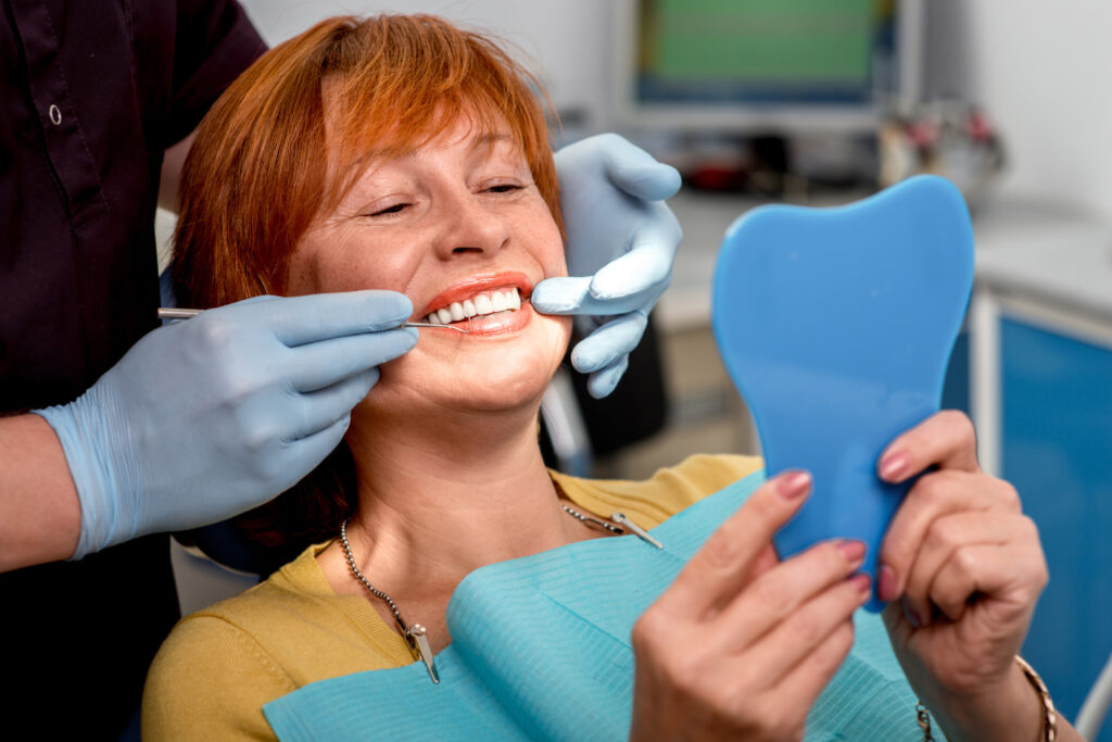
Implant-supported prostheses represent a significant advancement in restorative dentistry, utilizing titanium fixtures osseointegrated into alveolar bone to anchor dental restorations. Clinical studies demonstrate success rates exceeding 95% over ten-year periods when proper protocols are followed. These prosthetic solutions address edentulism through biomechanically stable frameworks that distribute occlusal forces effectively. Understanding the thorough treatment modalities, eligibility criteria, and long-term implications remains essential for ideal therapeutic outcomes.
Types of Implant-Supported Prostheses Available
When considering implant-supported prostheses, clinicians must evaluate three primary categories based on the extent of edentulism and specific patient requirements. Single-tooth implant crowns replace individual missing teeth, utilizing titanium or zirconia implant materials integrated with customized abutments. Fixed partial dentures, commonly termed implant bridges, span edentulous spaces by anchoring to multiple implants, eliminating the need for adjacent tooth preparation.
Complete-arch prosthesis designs include fixed hybrid prostheses and removable overdentures. Fixed hybrid prostheses employ four to eight implants supporting acrylic or zirconia superstructures through multi-unit abutments. Overdentures utilize two to four implants with locator or bar-clip retention systems, providing enhanced stability while permitting removal for maintenance. Selection criteria encompass bone volume, arch morphology, interocclusal distance, and patient-specific factors including manual dexterity and economic considerations.
How Dental Implants Work as Anchors for Prostheses
The biomechanical foundation of implant anchorage relies on osseointegration, a direct structural and functional connection between living bone tissue and the implant surface. Titanium implants undergo microscopic bonding with surrounding bone cells, creating stability equivalent to natural tooth roots. This biological fusion typically requires three to six months following surgical insertion.
Implant design considerations include thread geometry, surface topography, and diameter-to-length ratios, which influence primary stability and load distribution. Modern implants feature micro-textured surfaces that enhance cellular adhesion and accelerate osseointegration.
The implant placement technique determines prosthetic success through precise angulation and depth positioning. Clinicians utilize surgical guides and computed tomography data to achieve ideal bone engagement. Once integrated, implants transmit occlusal forces directly to surrounding bone, preserving skeletal architecture while supporting fixed or removable prosthetic components.
Benefits Over Traditional Dentures and Bridges
Beyond their osseointegrated foundation, implant-supported prostheses demonstrate measurable superiority over conventional dentures and bridges across multiple clinical parameters. Research indicates 95% five-year survival rates compared to 84% for fixed partial dentures. Implants preserve adjacent tooth structure, eliminating the 1.5mm reduction required for bridge abutments.
Biomechanically, implants restore 85-90% of natural chewing efficiency versus 25% with traditional dentures. The improved appearance results from customizable emergence profiles and elimination of visible clasps. Enhanced function manifests through increased bite force (200N versus 50N in complete dentures) and proprioceptive feedback via mechanoreceptors.
Bone preservation rates reach 0.2mm annual loss compared to 0.5-1.0mm with conventional prostheses. Speech articulation improves without palatal coverage. Psychological benefits include increased confidence and dietary variety, with 92% patient satisfaction rates documented in longitudinal studies.
The Complete Treatment Process and Timeline
The extensive treatment protocol for implant-supported prostheses spans multiple phases across 3-12 months, encompassing diagnostic evaluation, surgical placement, osseointegration, and prosthetic restoration. The initial planning phase requires CBCT imaging, diagnostic wax-ups, and surgical guide fabrication to establish precise implant positioning based on available bone volume and prosthetic requirements. Following surgical placement with primary stability of 35-45 Ncm, the osseointegration period necessitates 3-6 months for mandibular sites and 4-8 months for maxillary sites before proceeding to abutment connection and final prosthesis delivery.
Initial Planning Phase
How does successful implant-supported prosthetic rehabilitation begin? The initial planning phase establishes the foundation through extensive pre surgical planning protocols. Clinicians conduct thorough clinical examinations, evaluating bone density, soft tissue quality, and occlusal relationships. Diagnostic imaging, including cone-beam computed tomography (CBCT) scans, provides three-dimensional visualization of anatomical structures, enabling precise assessment of available bone volume and proximity to critical structures.
Digital treatment planning software facilitates virtual implant placement, determining ideal positioning, angulation, and dimensions. Radiographic templates and surgical guides are fabricated based on prosthetically-driven treatment plans. Interdisciplinary consultation between prosthodontists, oral surgeons, and laboratory technicians ensures coordinated care delivery. Risk assessment identifies potential complications, including inadequate bone support, sinus proximity, and nerve involvement. This systematic approach minimizes surgical complications and ensures favorable prosthetic outcomes.
Surgery to Restoration
Three distinct phases characterize the complete implant-supported prosthetic treatment timeline: surgical placement, osseointegration, and prosthetic restoration. Surgical techniques vary between immediate and delayed placement protocols, with each approach requiring specific implant placement considerations based on bone density, available vertical height, and proximity to anatomical structures. The surgical phase typically involves flap elevation, osteotomy preparation, and implant insertion at predetermined torque values of 35-45 Ncm.
Osseointegration requires 3-6 months, depending on bone quality and implant surface modifications. During this period, provisional restorations maintain function and aesthetics. The final prosthetic phase encompasses impression-taking, abutment selection, and crown fabrication using CAD/CAM technology or conventional laboratory methods. Treatment completion ranges from 4-9 months, with immediate loading protocols reducing timeline to 2-3 months in select cases meeting specific criteria.
Candidacy Requirements and Pre-Treatment Evaluation
Determination of patient suitability for implant-supported prostheses requires systematic evaluation through quantitative bone density measurements using cone-beam computed tomography to assess cortical thickness and trabecular architecture at proposed implant sites. Thorough medical history review identifies systemic conditions affecting osseointegration, including uncontrolled diabetes mellitus (HbA1c >7.5%), immunosuppression, bisphosphonate therapy, and active periodontal disease requiring stabilization prior to implant placement. Clinical and radiographic oral health examination establishes baseline periodontal status, occlusal relationships, and prosthetic space requirements while identifying pathological conditions necessitating pre-implant intervention.
Bone Density Assessment
While successful osseointegration fundamentally depends on adequate bone volume and quality at the recipient site, thorough radiographic and clinical assessment protocols must systematically evaluate trabecular architecture, cortical thickness, and overall bone density before implant placement. Cone-beam computed tomography provides three-dimensional visualization of alveolar bone morphology, enabling precise bone mass measurements through Hounsfield unit quantification. Clinicians classify bone quality using Lekholm-Zarb classifications (D1-D4), correlating density values with anticipated primary stability outcomes.
Skeletal health analysis incorporates dual-energy X-ray absorptiometry when systemic osteoporosis is suspected, particularly in postmenopausal women and elderly patients. Quantitative ultrasound techniques offer radiation-free alternatives for preliminary screening. Sites demonstrating inadequate density (below 350 HU) require augmentation procedures before implantation. Contemporary planning software integrates density mapping with virtual implant positioning, optimizing placement within zones of maximum cortical engagement and trabecular support.
Medical History Review
A detailed medical history evaluation establishes fundamental criteria for implant candidacy through systematic assessment of systemic conditions, pharmacological influences, and behavioral risk factors that directly impact osseointegration success rates. Clinicians must analyze patient medical history extensively, examining diabetes mellitus control (HbA1c levels), cardiovascular disease status, osteoporosis severity, and immunocompromising conditions. Bisphosphonate therapy, corticosteroid use, and anticoagulant medications require specific protocol modifications. Documentation of prior dental work reveals critical information regarding periodontal disease patterns, endodontic failures, and previous extraction complications that influence treatment planning. Tobacco use quantification, parafunctional habits, and radiation therapy to maxillofacial regions constitute absolute or relative contraindications. Allergic reactions to titanium or dental materials, though rare, necessitate investigation. This systematic review enables risk stratification and determines appropriate prophylactic measures, surgical modifications, and prosthetic design considerations essential for predictable outcomes.
Oral Health Examination
How does thorough oral health assessment determine implant candidacy beyond systemic medical considerations? Exhaustive intraoral examination evaluates periodontal status, measuring probing depths, clinical attachment levels, and bleeding indices to identify active disease. Radiographic analysis reveals bone density, height, and width at proposed implant sites through periapical and cone-beam computed tomography imaging. Clinicians assess existing dentition for caries, endodontic pathology, and occlusal dysfunction that may compromise implant stability.
Patient dental hygiene habits undergo scrutiny through plaque indices and gingival inflammation markers. Inadequate preventive oral care correlates with increased peri-implantitis risk, necessitating behavioral modification before treatment. Parafunctional habits, including bruxism and clenching, require documentation and management strategies. Soft tissue biotype classification determines surgical approach and aesthetic predictability. This multifactorial evaluation establishes baseline parameters for treatment planning and long-term prognosis.
Cost Breakdown and Insurance Coverage Options
Three primary cost components determine the total investment for implant-supported prostheses: surgical placement fees ranging from $1,500 to $3,000 per implant, abutment costs between $300 and $500 per unit, and prosthetic restoration expenses varying from $1,000 to $3,000 for single crowns or $3,500 to $30,000 for full-arch solutions. Additional expenses include diagnostic imaging ($100-$500), bone grafting procedures ($250-$3,000), and maintenance appointments ($200-$400 annually).
Most dental insurance plans classify implants as elective procedures, limiting coverage to 50% with annual maximums of $1,000-$2,000. Insurance pre-authorization remains essential for determining exact benefits. Medical insurance may provide coverage when tooth loss results from trauma or medical conditions. Cost comparison between traditional dentures and implant-supported alternatives reveals higher initial investment but superior long-term value through enhanced durability and reduced replacement frequency.
Long-Term Maintenance and Care Guidelines
While the financial investment in implant-supported prostheses represents a significant consideration, the longevity and performance of these restorations depend fundamentally on systematic maintenance protocols and patient compliance with care guidelines. Research demonstrates that adherence to periodic cleaning schedules correlates directly with implant survival rates exceeding 95% at ten-year intervals. Professional maintenance appointments should occur biannually, incorporating radiographic evaluation, torque verification of abutment screws, and peri-implant tissue assessment.
Proper cleaning techniques require specialized instrumentation, including plastic-tipped scalers and titanium-compatible curettes to prevent surface damage. Patients must implement twice-daily biofilm disruption using low-abrasive dentifrice, interdental brushes calibrated to embrasure dimensions, and antimicrobial rinses containing 0.12% chlorhexidine gluconate during acute inflammatory episodes. Documentation of probing depths, bleeding indices, and mobility scores enables early intervention when peri-implant pathology develops.
Potential Risks and Success Rates to Consider
Four primary categories of complications—biological, mechanical, iatrogenic, and patient-related—influence the clinical outcomes of implant-supported prostheses, with cumulative success rates ranging from 89.7% to 96.3% across contemporary longitudinal studies exceeding five years. Biological complications encompass peri-implantitis (prevalence: 10-15%), marginal bone loss exceeding 2mm, and soft tissue dehiscence. Mechanical failures include abutment screw loosening (8.8%), prosthetic fractures, and framework deformation under occlusal loading.
Surgical complications manifest as nerve paresthesia (0.5-3.7%), sinus membrane perforation, and hemorrhagic events. Early potential implant failure occurs through inadequate primary stability and infection, while late failures result from progressive bone loss and fatigue fractures. Patient-specific factors—uncontrolled diabetes (HbA1c >8%), active periodontitis, and smoking—significantly compromise osseointegration. Iatrogenic risks involve improper positioning, thermal necrosis during osteotomy, and contamination protocols. Meta-analyses demonstrate correlation between surgical experience and reduced complication rates.
Frequently Asked Questions
Can I Get an MRI Scan With Dental Implants in Place?
Patients with dental implants can safely undergo MRI scanning. Modern implant materials composition utilizing titanium or zirconia exhibits paramagnetic properties, causing minimal artifact. Post-implant placement procedure, these biocompatible alloys remain unaffected by magnetic fields during imaging.
Will Implant-Supported Prostheses Affect How Food Tastes?
Implant-supported prostheses typically preserve normal gustatory sensitivity since they don’t cover the palate extensively. Studies indicate minimal taste changes occur, with patients reporting comparable taste perception to natural dentition, unlike conventional dentures which obstruct taste receptors.
Can I Travel Immediately After Getting Dental Implants?
Patients should avoid air travel for 48-72 hours post-implantation due to barometric pressure changes affecting the initial healing period. Clinicians recommend gradual return to normal activities, with unrestricted travel typically permitted after seven to ten days.
Do Dental Implants Set off Metal Detectors at Airports?
Dental implants typically do not trigger airport metal detectors. The implant material composition, chiefly titanium or zirconia, exhibits low ferromagnetic properties. Modern implant detection sensitivity thresholds are calibrated to identify larger metallic masses, not small biocompatible fixtures.
Is Bone Grafting Always Necessary Before Implant Placement?
Bone grafting is not universally required. Clinicians determine necessity through bone density evaluation and radiographic assessment. Patients with adequate bone volume and sufficient alveolar ridge dimensions may proceed with immediate implant placement without augmentation procedures.