Dental X-rays play a pivotal role in modern dentistry by providing essential diagnostic information that is not visible during routine examinations. They enable the early detection of cavities, periodontal disease, and impacted teeth, which allows for timely intervention and treatment. By revealing hidden issues such as interproximal caries and bone loss, X-rays facilitate precise assessment and treatment planning. The advancements in imaging technology further enhance diagnostic accuracy while ensuring patient safety, setting the stage for improved oral health outcomes.
Understanding Dental X-Ray Technology
Dental X-rays are an essential diagnostic tool that employs electromagnetic radiation to capture images of the teeth and surrounding structures. This process, known as dental imaging, is pivotal for identifying oral health issues that are not visible during a routine examination. Radiographic techniques involve the use of X-ray machines that generate controlled, low-dose radiation to penetrate oral tissues. The resulting images provide detailed visual data, allowing clinicians to assess bone density, detect cavities, and evaluate the status of dental restorations. Modern advancements in digital radiography enhance image clarity and reduce radiation exposure, ensuring patient safety. Precision in radiographic techniques is vital for accurate diagnosis and treatment planning, making dental X-rays indispensable in contemporary dental practice.
Types of Dental X-Rays and Their Uses
Dental X-rays are categorized into various types, each serving specific diagnostic purposes. Bitewing X-rays are primarily utilized to visualize the crowns of the upper and lower teeth simultaneously, aiding in the detection of cavities and evaluating bone density. In contrast, panoramic X-rays provide an extensive view of the entire oral cavity, offering valuable insights into jaw structure, tooth positioning, and identifying potential pathologies.
Bitewing X-Rays Purpose
Bitewing X-rays serve as a critical diagnostic tool within dentistry, providing detailed images that reveal the upper and lower teeth in a single view. They are instrumental in the early detection and monitoring of interproximal cavities, making them an invaluable resource for dental professionals. The bitewing benefits extend to examining bone levels and detecting periodontal disease by revealing the alignment and spacing of teeth. Their precision allows for accurate cavity detection, especially in areas not visible through a standard oral examination. This capability enables timely intervention, reducing the risk of extensive decay. Additionally, bitewing X-rays assist in evaluating existing restorations, ensuring their integrity and fit. Overall, bitewing X-rays enhance diagnostic accuracy, contributing notably to preventive dental care.
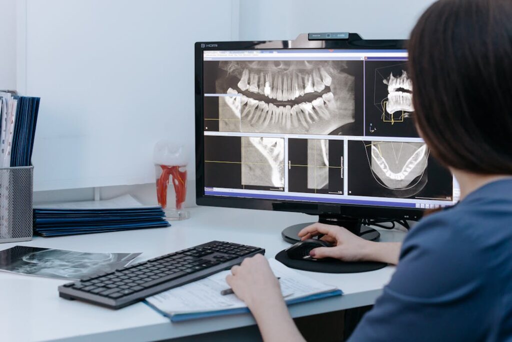
Panoramic X-Rays Benefits
Panoramic X-rays offer a detailed view of the entire oral cavity, providing a single, wide-angle image that encompasses teeth, jaws, and surrounding structures. This technique, known as panoramic imaging, is vital in diagnosing a variety of dental and skeletal issues. It allows for the assessment of jaw alignment, which is essential for detecting temporomandibular joint disorders and planning orthodontic treatments.
Furthermore, panoramic X-rays are important for identifying impacted teeth, cysts, tumors, and other anomalies that might not be visible through standard intraoral X-rays. They provide valuable insights into the overall health of the oral cavity with minimal exposure to radiation. As a diagnostic tool, panoramic X-rays considerably enhance the clinician’s ability to formulate extensive treatment plans and guarantee ideal patient outcomes.
The Role of Dental X-Rays in Detecting Cavities
Although often unnoticed by the naked eye, cavities can be effectively identified through the precise application of dental X-rays. This imaging modality is essential among cavity detection techniques, providing a detailed view of the tooth’s internal structure. X-rays enable clinicians to detect carious lesions at an incipient stage, which is critical for preventing further decay and preserving dental integrity. The early diagnosis benefits afforded by X-rays include timely intervention and minimally invasive treatments, which can mitigate the need for extensive restorative procedures. By revealing interproximal caries and sub-surface enamel erosion, dental X-rays allow for a thorough assessment that visual inspection alone cannot provide. This enhances the accuracy of diagnoses, ensuring patients receive appropriate and effective care promptly.
Identifying Impacted Teeth With X-Rays
Dental X-rays are essential for identifying impacted teeth, as they reveal issues not visible during a standard oral examination. By providing detailed images of tooth positioning, X-rays allow practitioners to assess potential complications such as misalignment or infection risk. This information is vital for planning effective treatment strategies, ensuring ideal patient outcomes.
Detecting Hidden Tooth Issues
Radiographs serve as an essential tool in the detection of hidden dental issues, particularly when identifying impacted teeth. Their ability to penetrate oral structures allows for the visualization of hidden cavities and underlying tooth decay that are not perceptible during a standard visual examination. Impacted teeth, often concealed beneath the gum line, can lead to significant oral complications if left undiagnosed. X-rays provide a vital perspective on the orientation and position of these teeth, enabling a precise assessment of their potential interference with adjacent teeth. This diagnostic capability guarantees early intervention, preventing the progression of dental conditions that could otherwise compromise oral health. Through the careful interpretation of radiographic images, dental practitioners can devise effective treatment plans tailored to the patient’s specific needs.
Assessing Potential Complications
How effectively can potential complications be evaluated with the use of X-rays in identifying impacted teeth? Dental X-rays serve as an essential diagnostic tool, providing thorough insights into the positioning and condition of teeth. By capturing detailed images, X-rays facilitate the identification of impacted teeth, which are teeth that have failed to emerge properly due to obstructions or malalignment. This imaging technique is instrumental in evaluating growth patterns that may indicate potential impaction. Additionally, X-rays are critical in identifying infections that could arise from impacted teeth, such as abscesses or cysts. Such complications, if unaddressed, could lead to more severe oral health issues. Consequently, X-rays are indispensable for dental professionals in preemptively evaluating potential risks associated with impacted teeth.
Planning Effective Treatments
Identifying impacted teeth with X-rays not only aids in evaluating potential complications but also plays a significant role in planning effective treatments. Through precise imaging, dental professionals can accurately assess the position and condition of impacted teeth, facilitating the development of tailored treatment strategies. These strategies often include surgical intervention or orthodontic solutions, depending on the specific needs of the patient. X-rays provide vital data that enable clinicians to anticipate possible challenges and devise a thorough plan, thereby optimizing outcomes. In addition, patient collaboration is essential; sharing X-ray findings with patients fosters informed decision-making and enhances compliance with recommended treatments. The integration of advanced imaging technologies guarantees a meticulous approach to managing impacted teeth, ultimately contributing to improved oral health.
Assessing Bone Health and Detecting Bone Loss
When evaluating oral health, dental X-rays are instrumental in appraising bone health and detecting bone loss. Through precise x-ray interpretation, clinicians can assess bone density, which is essential for diagnosing various conditions such as osteoporosis and periodontal disease. X-rays provide a detailed view of the alveolar bone supporting the teeth, allowing practitioners to identify even subtle changes in bone structure. The ability to detect bone loss at an early stage is critical for implementing timely interventions, thereby preventing further complications. Additionally, X-rays enable the monitoring of progression in degenerative conditions, facilitating informed clinical decisions. By offering a non-invasive method to visualize internal structures, dental X-rays contribute greatly to the thorough evaluation of a patient’s oral health status.
Enhancing Treatment Planning and Precision
Building on the insights gained from evaluating bone health, dental X-rays play a pivotal role in enhancing treatment planning and precision. By providing detailed images of the dental and periodontal structures, X-rays considerably improve treatment accuracy. Clinicians are empowered to develop thorough treatment strategies that address specific patient needs. The precision afforded by dental X-rays facilitates the identification of underlying issues such as root canal infections, impacted teeth, and orthodontic anomalies. These insights enable practitioners to tailor interventions that optimize patient outcomes. Additionally, the ability to visualize the spatial relationship of anatomical structures ensures that restorative procedures are executed with high accuracy. To summarize, dental X-rays are indispensable tools that enhance the clinician’s ability to deliver precise and effective oral healthcare.
Safety Measures and Advancements in X-Ray Technology
As dental X-ray technology continues to evolve, significant strides have been made in enhancing safety measures for both patients and practitioners. Radiation safety remains a vital priority, with modern X-ray systems designed to minimize exposure while maintaining diagnostic efficacy. State-of-the-art imaging advancements, such as digital radiography, utilize lower radiation doses compared to traditional film-based methods. These advancements include the introduction of collimators, which focus the X-ray beam, reducing scatter radiation. Additionally, the implementation of lead aprons and thyroid collars further protects patients from unnecessary exposure. For practitioners, adherence to the ALARA (As Low As Reasonably Achievable) principle guarantees that radiation exposure is kept to a minimum. Collectively, these measures underscore the commitment to safe and effective dental imaging.
The Importance of Regular Dental X-Rays
Regular dental X-rays are a critical component in maintaining oral health, providing an extensive view of teeth and supporting structures that are not visible during a routine examination. The importance of prevention is underscored by the ability of X-rays to identify early signs of dental issues such as caries, periodontal disease, and abscesses. By detecting these problems early, dental professionals can implement timely interventions, reducing the risk of more complex and costly treatments. Moreover, patient education is enhanced through the use of X-rays, as they offer a visual tool to help patients understand their oral health status and the necessity for preventive actions. Regular X-rays are indispensable in devising detailed treatment plans and fostering informed decisions for ideal oral care.
Future Developments in Dental Imaging
Advancements in dental imaging are poised to revolutionize oral health diagnostics by integrating cutting-edge technology with traditional practices. Emerging imaging techniques, such as cone beam computed tomography (CBCT) and digital radiography, offer enhanced precision and reduced exposure to radiation. These techniques provide detailed 3D representations of dental structures, facilitating superior diagnostic accuracy. AI advancements further augment these capabilities by enabling automated interpretation of complex datasets, enhancing the detection of pathologies with improved speed and reliability. AI-driven algorithms can analyze vast amounts of imaging data, identifying patterns imperceptible to human practitioners, thereby aiding in early disease detection and treatment planning. Future developments will likely focus on refining these imaging techniques and AI integration to optimize patient outcomes in oral health diagnostics.
Frequently Asked Questions
How Often Should Children Get Dental X-Rays?
The recommended x-ray frequency for children’s dental health varies based on individual risk factors, including age, oral hygiene, and history of dental issues. Generally, dentists assess these factors to determine ideal intervals for diagnostic imaging practices.
Do Dental X-Rays Show Signs of Oral Cancer?
Dental x-rays provide essential benefits in oral cancer detection by revealing anomalies in hard tissues. While x-rays mainly detect bone changes, they can assist in identifying potential signs warranting further investigation for oral cancer diagnosis.
Can Dental X-Rays Detect Gum Disease?
Dental x-rays can detect gum disease by revealing bone loss, a key gum disease symptom. X-ray benefits include visualizing areas not visible during a clinical exam, providing critical information for accurate diagnosis and treatment planning of periodontal issues.
Are There Any Alternatives to Traditional Dental X-Rays?
Alternatives to traditional dental X-rays include digital imaging and ultrasound technology. Digital imaging allows for enhanced image quality and reduced radiation exposure, while ultrasound technology provides non-invasive tissue analysis, offering a thorough approach to oral health evaluation.
How Do Dental X-Rays Differ From Other Medical X-Rays?
Dental x-rays differ from other medical x-rays primarily in film types and exposure levels. Dental x-rays utilize specialized films that require lower exposure levels, focusing on high-resolution images of teeth and gums for accurate oral health assessment.